Friday, January 8, 2021

Lupine Publishers | Infra-red Radiation in Diagnostic of Arteriogenic Erectile Dysfunction

 Lupine publishers | Journal of Urology & Nephrology Studies


Goal: To identify influence of long range infra-red radiations on endothelium- dependent vasodilatation in patients with erectile disorders.

Material and Methods: One hundred eighteen males with erectile disorders were examined. Twenty practically healthy males comprised a control group. An endothelial function of the cavernoma arteries was determined with ultrasonic Diplography after of application of a long range infra-red radiation.

Results: An endothelial dysfunction of the cavernous arteries revealed with a Drug-Diplography method in the main group was also proved at the use of the original technique. Paradoxical vasoconstriction which was found during utilization of drug-induction method assay was identified in some patients. A direct correlation between these techniques was revealed and the correlation ratio was r=0.58 (p<0.05).

Conclusion: The method developed is informative in diagnostics of endothelial dysfunction of the cavernoma arteries, and we think that it will take a relevant place in a comprehensive examination of patients with erectile disorders.

Keywords: long range infra-red radiation; erectile dysfunction; endothelial dysfunction.

Introduction

The main diagnostic method for vasculogenic erectile dysfunction (ED) at the percentage is diplography of the cavernous arteries of the penis after intracavernous administration of vasoactive drugs- pharmacodopplerography (FDG) [1]. This method has several disadvantages-pain and fear of the patient before injection into the penis, which can reduce the diagnostic value of the results [2]. Some of these shortcomings are deprived of the technique of dopplerography of the vessels of the penis after taking 5-phosphodiesterase inhibitors [3]. However, the results of this study are more dependent on the degree of sexual arousal of the patient. It should be noted the high cost of such a study [4], and the duration (in time) of the survey. Decreased abilities of the endothelium of the arteries and sinuses of the penis to synthesize and secrete nitric oxide (NO) lead to impaired relaxation of smooth muscle cells of the vascular wall, which limits blood flow and prevents the development of an erection [5,6].
Widely used in the diagnosis of endothelial dysfunction of peripheral arteries, the method of ultrasound examination of postcompression changes in their diameter [6]. The degree of expansion of peripheral arteries reflects the ability of endothelial cells to synthesize and secrete NO and its bioavailability, and, consequently, the state of endothelial function [6]. The mechanism of post-occlusal changes in the diameter of blood vessels is currently explained by a sharp increase in blood flow in the arteries due to the expansion of the distal small vessels and the mechanical effect of increased blood flow on the vascular wall, which leads to the activation of ion channels of endothelial cells and the accumulation of calcium ions in them. The latter activate endothelial NO synthetase, which is accompanied by the release of NO, which has a vasodilating effect on the vascular wall.
R. Virag [7] applied the technique of post-occlusal changes in the diameter of peripheral arteries to detect endothelial dysfunction in the pool of cavernous arteries, which allows differentiating different pathogenetic forms of ED at the stage of functional disorders. The sensitivity and specificity of this indicator is 100 and 92%, respectively. An improved methodology for the post-compression test in the diagnosis of vasculogenic erectile dysfunction was proposed by Maso EB et al. [8]. The sensitivity and specificity of the proposed methodology are 100 and 94.5%, respectively. However, the technique has several drawbacks-a certain technical complexities of execution, the presence of discomfort in the area of the cuff during compression.
It is known that hemoglobin NO complexes are photosensitive and decompose with the release of nitric oxide. The photorelaxing effect of infrared (IR) radiation on blood vessels is well known. At present, a positive effect of long-range infrared radiation on the functional properties of platelets and rheological parameters during irradiation of blood of patients with angina pectoris in vitro has been identified [9-11]. Infrared radiation in the terahertz range causes an increase in the production of nitric oxide by vascular endothelium, which is accompanied by the normalization of reduced basal and induced vasodilating activity in animals in a state of acute immobilization stress [12]. According to some modern authors, when irradiating the infrared radiation of the terahertz range, not only can the synthesis of endogenous nitric oxide and its reactivity increase, but the duration of the existence of nitric oxide in cells can also increase [13].
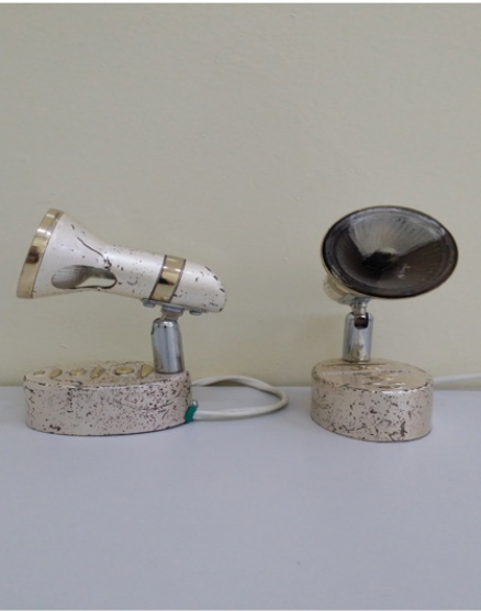
The purpose of this study is to assess the effect of long-range infrared radiation on endothelium-dependent vasodilation in patients with erectile dysfunction. Materials and methods. The study included 118 men aged 32-71 years with ED, as well as 20 men (control group) aged 28-66 years without ED. All men with ED underwent a comprehensive examination, which included the collection of a general medical and sexological history, a survey on the International Index of Erectile Function, a general examination, a study of hormonal status, blood lipids and glucose, as well as FGD using vasoactive drugs (papaverine 20-40 mg; alprostadil 5-10mkg). In addition, the study of the endothelial function of the cavernous arteries was performed in all patients. The function of the arterial endothelium was evaluated using ultrasonic examination (ultrasound) of the diameter before and after the use of long-range IR emitters (Figure 1). (ZB series registration certificate No. UZTT 00898 - working wavelength range of useful radiation 22.5 μm) according to our developed methodology. The experimental nature of the test was explained to all participants and consent was received from them to conduct it. Ultrasound of the cavernous arteries was performed using an “HD3” device from Philips (Holland).
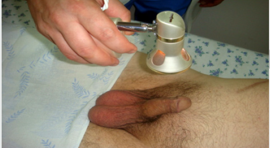
In the position of the patient on the back, a linear sensor L 9-5 was placed longitudinally on the ventral surface of the penis. The diameter of the cavernous arteries was measured as the distance between the opposite walls of the vessel and the location of the sensor was noted with a pencil. After determining the average diameter of the cavernous arteries of the penis, an IR emitter (ZB series) was exposed with an exposure of 3 minutes from the ventral side at a distance of 10-12 cm from the penis (Figure 2). After 3 minutes, an ultrasound scan was repeated to measure the diameter of the cavernous arteries. For the calculation, the largest diameter of the cavernous arteries was used. The percentage of increase in the diameter of the cavernous artery (PUDKA) was adopted by us as the main indicator for assessing the endothelial function of the cavernous arteries. This indicator was calculated by the formula:

Figure 1: Far-range infrared emitters.

lupinepublishers-openaccess-journal-urology-nephrology

Figure 2: Methods of exposure of IR emitter to cavernous arteries

lupinepublishers-openaccess-journal-urology-nephrology

PUDKA = 100% x (D2 - D1) / D1,
where D1 is the diameter of the cavernous artery before exposure to an infrared emitter;
D2 - diameter of the cavernous artery after exposure to an infrared emitter.

All studies were carried out by one specialist in ultrasound diagnostics. The study was carried out in the morning, before the study, patients were advised to refrain from smoking and taking medications that affect the cardiovascular system. The average values of this indicator were calculated for the control group and patients with arterial and non-arterial ED. Statistical analysis was performed using t-student criterion and Pearson correlation coefficient. For analysis, computer software Statistica 6.0 was used; p <0.05 was recognized as statistically significant.

Results

divided into 2 subgroups: subgroup I - non-arterial ED of 42 patients (peak systolic velocity of 30 cm / s or more); Subgroup II - arteriogenic ED of 76 patients (peak systolic velocity less than 30 cm \ s). A comparative analysis of the average PUDA values in patients with different forms of ED revealed that this indicator is statistically significantly (p <0.001) less in the group of patients with arteriogenic ED compared with the control group, as well as in comparison with patients with other forms ED. An analysis of the data made it possible for us to propose a threshold level of PUDA for distinguishing organic ED from other forms of erectile disturbances, which amounted to 30%. The sensitivity and specificity of this indicator relative to the results of FDG in the diagnosis of arteriogenic ED were 100% and 96%, respectively. In patients with arteriogenic ED there was a significant correlation (r = 0.58 p <0.05) with the results of FDG (peak systolic rate) and PUDK values. An analysis of the results showed that the proposed method reveals a paradoxical vasoconstriction of the cavernous arteries, which is not determined when using FDG. When conducting the proposed diagnostic method, in no case did we observe complications and adverse reactions.

Discussion

The present study shows that the use of long-range IR emitters in determining the endothelial function of the cavernous arteries using ultrasound is a highly informative method for the diagnosis of arteriogenic ED. The developed technique for studying the endothelial function of the cavernous arteries has advantages over FDG and post-occlusion test, as it is a non-invasive method and technically simple to perform. It is known that the functional state of penile arteries depends on the biological activity of NO. Endothelial cells and nerve endings of non-cholinergic nonadrenergic neurons are sources of NO in the cavernous tissue of the penis. The synthesis of NO is carried out as a result of the action of enzymes of endothelial and neuronal NO synthase [14,15]. In our opinion, endothelial and neuronal NO synthases are activated by the quantum energy of the IR emitter, which leads to the release of NO. It is known that the quantum energy of radiation is inversely proportional to the wavelength, and if you take this into account, the effect on a person with radiation in the range of 8-9.8 μm (human own radiation is in the range of 9-10 μm) does not have negative side effects, since quantum energy of such sources does not exceed the quantum energy of the skin’s own radiation. The radiation of ceramic infrared emitters has a specific wavelength in the narrow spectral range. All types of emitters are characterized in that the energy spectrum of their exposure corresponds to or below the energy spectrum of human radiation. Emitters in these ranges have no effect on a healthy person, since it is transparent to them [16,17]. The paradoxical vasoconstriction detected by the proposed method is due to the presence of oxidative stress. Longrange infrared radiation stimulates the production of superoxide radicals (NO; O) by blood phagocytes and tissue macrophages, which enhances oxidative stress. This is not observed during FDG. Endothelial dysfunction is an early stage of atherosclerosis and is systemic in nature. Functional damage to most arterial vessels often proceeds without clinical symptoms; therefore, the timely detection of early stages of vascular system damage is relevant. In our opinion, the developed method for determining the endothelial function of the cavernous arteries is able to detect endothelial dysfunction at the level of functional disorders, which is not always determined with FDG.

Conclusion

Thus, our proposed method for studying the endothelial function of the cavernous arteries after exposure to a far-infrared emitter is highly informative in the diagnosis of arteriogenic ED, especially in the stage of functional disorders. This method will take its place in a comprehensive examination of patients with ED. Conflict of interest. The author claims no conflict of interest.

Acknowledgment

To the Head of the Clinical and Diagnostic Department of the Central Clinical Hospital No. 1 of the Main Medical Directorate under the Office of the President of the Republic of Uzbekistan, Doctor of Medical Sciences G. A. Rozykhojaeva.

https://twitter.com/lupine_online


Radiology; USG and Colour Doppler of Post Renal Transplant Complications

  Abstract Kidney transplant is the treatment of choice for patients with end-stage renal disease. Kidney transplant offers better...