Showing posts with label lupine publishers journals. Show all posts

Wednesday, September 20, 2023

Radiology; USG and Colour Doppler of Post Renal Transplant Complications


Abstract

Kidney transplant is the treatment of choice for patients with end-stage renal disease. Kidney transplant offers better quality of life. It is more cost effective than hemodialysis. Advances in surgical technique, along with improvement in organ preservation and immunosuppression have improved patient outcomes. Post-operative complications, however, can limit this success. Ultrasound and Doppler study is the primary imaging modality for evaluation of renal transplant, providing real –time information about complication in graft. A multimodality approach including CT scan, MRI or conventional angiography may be necessary in cases when sonography and Doppler are inconclusive to diagnose the etiologies of these complications. Radiologists play an integral role within the multidisciplinary team in care of transplant patient at every stage of the transplant process. Therefore, the radiologist should always be aware when evaluating the failing renal graft, whether the cause is renal or extrinsic. In this pictorial essay we tried to gather the most common complication of transplant kidney in different cases that occurred in our hospital, with an emphasis on Ultrasound and Doppler study.


Read more about this article: https://lupinepublishers.com/urology-nephrology-journal/fulltext/radiology-usg-and-colour-doppler-of-post-renal-transplant-complications.ID.000164.php

Read about Lupine Publishers Google Scholar Articles: https://scholar.google.com/citations?view_op=view_citation&hl=en&user=XbnlB50AAAAJ&cstart=400&pagesize=100&citation_for_view=XbnlB50AAAAJ:Nufq_to8ts0C


Monday, August 28, 2023

Evaluation of Buccal Mucosal Graft Urethroplasty for The Treatment of Female Urethral Strictures- A Single Centre Experience


Abstract

Introduction: Female urethral stricture is a highly under-reported and underdiagnosed condition encountered by the reconstructive urologist. Urethral dilatation is often performed with urethroplasty offered in select cases. In the present study, we describe our results in a series of women surgically treated for female urethral stricture disease using a suprameatal approach with a buccal mucosal graft dorsal on lay technique.
Materials and Methods: All females diagnosed of urethral stricture who underwent buccal mucosal graft urethroplasty from January 2015 to January 2020 were evaluated retrospectively. Intraoperative and postoperative parameters were assessed.
Results: A total of 14 female patients underwent buccal mucosal urethroplasty were evaluated. The mean age of the patients was 49.5 years ranging from 35 to 64 years. Mean preoperative maximum flow rate [Qmax] on uroflometry was 6.5 ml/second and the mean residual urine 156 ml. All patients underwent uneventful buccal mucosal graft dorsal on lay technique. At 3 months follow up, the mean Qmax was 23.2 ml/second with mean residual urine of 14 ml. A Self-reporting satisfaction scores using the Patient Global Impression of Improvement showed that seven patients scored 1 (very much better), four scored 2 (much better), two patients scored 3 (a little better), and one scored 4 (no change) none of the patients scored a 5 (worse).No recurrence was noted.
Conclusion: Buccal mucosal graft urethroplasty is a feasible surgery for female urethral strictures with minimal short term complications.


Read more about this article: https://lupinepublishers.com/urology-nephrology-journal/fulltext/evaluation-of-buccal-mucosal-graft-urethroplasty-for-the-treatment-of-female-urethral-strictures-a-single-centre-experience.ID.000163.php

Read about Lupine Publishers Google Scholar Articles: https://scholar.google.com/citations?view_op=view_citation&hl=en&user=XbnlB50AAAAJ&cstart=400&pagesize=100&citation_for_view=XbnlB50AAAAJ:DIubQTN3OvUC

Tuesday, May 16, 2023

Reasons and Outcome of Patients after Permanent Transfer from Peritoneal Dialysis to Hemodialysis: A Review of 16 years of Experience in Senegal


Abstract

Introduction: Peritoneal dialysis (PD) and haemodialysis (HD) are two complementary and non-competitive renal replacement therapy (RRT). A patient can be transferred from one technique to the other. The objective of this study was to assess the reasons for transferring patients from PD to HD and to follow their outcome.
Patients and Methods: This is a 16-year descriptive and analytical retrospective study (March 1, 2004 - August 31, 2020) conducted at the PD unit of the Aristide Le Dantec University Hospital in Dakar. Were included, patients on PD for at least 30 days, over 18 years of age and permanently transferred to HD. The probability of survival for any duration of post-transfer follow-up was estimated by the Kaplan-Meier method.
Results: The analysis covered 98 out of 113 cases. The mean age of the patients was 45.2 ± 14.09 years at the initiation of PD and 47 ± 13.91 years at the time of transfer, with a sex ratio of 0.66. The mean duration in PD was 19.9 ± 17.25 months [range, 1.0-90.0 months]. The transfer to HD concerned 73.5% of patients in the first two years. The reasons for transfer were mainly associated with infection (82.7%), mechanical complications (23.5%), social reasons (12.2%) and inadequate dialysis (6.1%). It was programmed in 11.4% of cases and 6% of patients had a permanent approach. At the endpoint date, the mean duration in hemodialysis was 43.3 months with 42.8% of patients still in HD. There was a kidney transplant patient; a return to PD. Mortality was 34.6%. The mean HD survival was 126 months. There was a statistically significant relationship between infection as a reason for transfer and mortality (p = 0.047).
Conclusion: The main reasons for transferring PD to hemodialysis identified in the literature are found in our context. This transfer must be anticipated to reduce morbidity and mortality.

Read more about this article: https://lupinepublishers.com/urology-nephrology-journal/fulltext/reasons-and-outcome-of-patients-after-permanent-transfer-from-peritoneal-dialysis-to-hemodialysis-a-review-of-16-years-of-experience-in-senegal.ID.000161.php

Read about Lupine Publishers Google Scholar Articles: https://scholar.google.com/citations?view_op=view_citation&hl=en&user=XbnlB50AAAAJ&cstart=400&pagesize=100&citation_for_view=XbnlB50AAAAJ:oYLFIHfuHKwC

Monday, April 10, 2023

A Rare Case of Penile Schwannomatosis Presenting with Painful Nocturnal Penile Tumescence


Background: Penile schwannoma is a rare tumor. They commonly present as an asymptomatic, painless and slow growing mass. Other presentations include sexual dysfunction, most commonly dyspareunia, followed by erectile dysfunction, abnormal penile curvature or pain with ejaculation.
Case presentation: A 26-year-old male presented atypically with painful nocturnal penile tumescence, along with multiple nodules over the dorsal penis. Excision of multiple penile tumors under general anaesthesia was performed and histopathologic examination revealed benign schwannoma.
Conclusion: Our hypothesis is that the schwannoma lies along the axis of the dorsal penile nerve, and compression of this nerve occurs during his erection causing pain. However, there are limited presentations of painful erections in penile schwannomas, and we hope that future studies can help confirm this theory.

Read more about this article: https://lupinepublishers.com/urology-nephrology-journal/fulltext/a-rare-case-of-penile-schwannomatosis-presenting-with-painful-nocturnal-penile-tumescence.ID.000160.php

Sunday, February 26, 2023

Swathed Ureter, an Enigma in Diagnosis- A Pictorial Essay

  Abstract

This pictorial essay is an educational article aiming to provide both textual and visual portrayals consisting of a collection of images and texts on an important issue by reviewing and extrinsic encasing pathology of the ureter to provide a guide to those who are involved in diagnostic intervention. While considering diseases of the urinary system, physicians mainly focus on the kidneys and the bladder. Only scant attention is paid to the ureters. Most of the UTO due to calculi are readily identifiable whereas many cases of ureteric exterior encasement are frequently missed from early detection even by experienced clinicians and radiologists. Failure in recognition of the encasement of ureters and its causes may lead to mistaken diagnosis with resultant inappropriate management. However, problems with the ureters can adversely affect the functioning of the kidneys and could even be lethal. In this article we focus only ‘encasement of ureters’ with a few common examples and salient signs that help in the diagnosis.

Read more about this article: https://lupinepublishers.com/urology-nephrology-journal/fulltext/swathed-ureter-an-enigma-in-diagnosis-a-pictorial-essay.ID.000158.php

Read about Lupine Publishers Google Scholar Articles: https://scholar.google.com/citations?view_op=view_citation&hl=en&user=XbnlB50AAAAJ&cstart=400&pagesize=100&citation_for_view=XbnlB50AAAAJ:cSdaV2aYdYsC


Sunday, January 29, 2023

Lasers in Urology – what has Survived of our Research Starting 1970*


Short Communication

In 1970 I started to investigate the Laser-Technology for urologic surgery together with H. Müßiggang. First of all, it was to check the interacting of the different lasers with biological tissue. The result you can gather from the Figure 1 Decisive are the two factors: absorption and scattering of light into the tissue. The strong light absorption of the CO2 – laser leads to an excellent incision effect with low edema reaction. The light of the is mainly absorbed in the tissue by hemoglobin and pigment colorings and therefore suitable for the destruction of highly vascularized tumors or malformations. For achieving greater volume effects, - necessary for destruction of solid tumors, bilharzial bladder-lesions and inflamed areas in interstitial cystitis, - the Nd:YAG – laser was used by us since 1976 (Figure 2). Presupposed for the clinical application of lasers was the developing of a quartz glass fiber transmission system by Nath, a physicist from the Neuherberg Laser Labor (1973) and the developing of a special cystoscope insert, designed by my working-group (Staehler, Frank et al.) and constructed by the Storz Compagny/Tuttlingen/Germany (Figure 3). The next steps were the laser induced shock wave lithotripsy, developed between 1978 to 1986 (Munich/Lübeck) and the photodynamic procedures for early tumor diagnosis (Figures 5a & 5b).

Read more about this article: https://lupinepublishers.com/urology-nephrology-journal/fulltext/lasers-in-urology-what-has-survived-of-our-research-starting-1970.ID.000157.php

Read about Lupine Publishers Google Scholar Articles: https://scholar.google.com/citations?view_op=view_citation&hl=en&user=XbnlB50AAAAJ&cstart=400&pagesize=100&citation_for_view=XbnlB50AAAAJ:K4-iKlO5MD4C

Tuesday, January 10, 2023

Chronic Kidney Disease, Data from MIKD


Abstract

Chronic kidney disease (CKD) is a leaping up public health matter. It has an augmented effect on cardiovascular diseases and affects every system in body. In developed countries the extent of prevalence is available due to renal registries. In middle or low socio economic countries the proportion of this evolving health issue is not known. CKD is rising and a major contributor is soaring number of diabetes mellitus worldwide. Other significant addition in this cohort of CKD is by rising numbers of obese and hypertensive patients. In order to curtail this rising health matter immediate and intense measures both at preventive and curative levels are required.

Aim: Due to lack of formal renal registry we wanted to see spectrum of chronic kidney disease (CKD) in our catchment area and at what CKD stage they present to tertiary care hospital.

Methods: All patients who presented to Emergency department of Multan Institute of kidney disease from 01 Sep 2017 till Sep 2019 data were evaluated. Record of Emergency department patients were taken from electronic system of our hospital. Some patients had multiple visits and we took first visits kidney function in our analysis. eGFR was calculated from serum creatinine with help of CKD-EPI equation.

Results: Total 4303 patients were included in study. Males were 60% and females 40%. Age range from 13 years to 96 year old. 945 patients were excluded as they were not falling in chronic kidney disease category. Remaining 3358 patients had chronic kidney disease. Sub-analysis according to CKD stage showed 66.17% patients presented at CKD stage V.

Conclusion: Kidney disease is rising globally. Countries where renal registries are established provide incidence of chronic kidney disease ranging from 10 to 15%. Still a lot of countries worldwide do not have established system of data collection so true incidence is not established. Our work is first of its kind reported from this area. Drastic preventive strategies are need of time from health budget planners.

Read more about this article: https://lupinepublishers.com/urology-nephrology-journal/fulltext/chronic-kidney-disease-data-from-mikd.ID.000156.php

Read about Lupine Publishers Google Scholar Articles: https://scholar.google.com/citations?view_op=view_citation&hl=en&user=XbnlB50AAAAJ&cstart=400&pagesize=100&citation_for_view=XbnlB50AAAAJ:BW2nPTmhBn4C


Monday, December 5, 2022

Efficacy and Safety of Prolonged Alfuzosin Treatment in Patients with Lower Urinary Tract Symptoms Associated with Benign Prostatic Hyperplasia: 7 Years of Observation

Abstract

Introduction: Amongst all of the medications prescribed to BPH patients undergoing conservative therapy, α1-blockers were used in 80% of cases. The question of optimal treatment length is one that has been constantly asked during the last years. The current study will encompass the data we have collected of a small study group of BPH patients treated with 10 mg of Alfuzosin daily, over a period of more than 7 years.

Material and Methods: From 2009 to 2011, 41 patients with mean age 67.7 years began medical therapy taking 10 mg of Alfuzosin per day. The mean treatment length was 7.6 years as of today. 16 patients (39%) with prostates exceeding 60 cm3 were additionally prescribed 5-ARIs.

Results: Overall, a positive dynamic was found in 85.4% of patients. Not a single patient chose to discontinue treatment. A very high level of satisfaction was reported by 88% of our patients. A statistically significant (P < 0.05) decrease in IPSS scores by 8.5 ± 6.1 (47.5%) was found. Mean QoL indices decreased from 3.7 ± 1.1 to 2.3 ± 1.1 over the period of observation. Mean Qmax values increased from 9.7 ± 0.52 mL/s to 14 ± 0.60 mL/s (an increase of 44.3%).

Conclusions: This study has demonstrated a high level of safety and efficacy when using Alfuzosin to treat voiding dysfunction in patients suffering from LUTS/BPH. Our study resolves the issue surrounding a prolonged course of α1-blockers, which, due to its widespread use, remains the gold standard for medical treatment of lower urinary tract symptoms associated with benign prostatic hyperplasia.

Read more about this article: https://lupinepublishers.com/urology-nephrology-journal/fulltext/efficacy-and-safety-of-prolonged-alfuzosin-treatment-in-patients-with-lower-urinary.ID.000154.php

Monday, November 28, 2022

Robot-Assisted Radical Prostatectomy our Technique Description


Abstract

Objective: To describe step-by-step technique in Robot Assisted Radical Prostatectomy of our transperitoneal posterioranterior technique for prostatic dissection with preservation of the endopelvic fascia, preservation of the puboprostatic ligaments and dorsal venous complex.

Materials and Methods: Description of our surgical technique over 80 patients who underwent RARP and the characteristics group from 2016 to 2019 excluding the rest of cases who went to different surgical approach and technique for robot-assisted radical prostatectomy.

Results: The mean age was 63 years old, 7% of patients were overweight and 7.5% had obesity. The mean pre-operative prostate volume was 42.620 cc, mean prostatic specific antigen (PSA) of 10.414.8 ng/dl. The mean console time was 198±47. The surgical margins were positive in 13.75% of the patients. Complications were recorded in the peri-operative period, five (6.2%) Clavien-Dindo I and six (7.5%) Clavien-Dindo II.

Conclusions: After 8 years of experience in our center we have modified our technique of robot assisted radical prostatectomy, improving our results, following different worldwide concepts in the prostatic dissection. Even if necessary, to increase the number of cases we have find an easier way to reproduce with acceptable results.


Read more about this article: https://lupinepublishers.com/urology-nephrology-journal/fulltext/robot-assisted-radical-prostatectomy-our-technique-description.ID.000153.php

Read about Lupine Publishers Google Scholar Articles: https://scholar.google.com/citations?view_op=view_citation&hl=en&user=1s6IPw0AAAAJ&citation_for_view=1s6IPw0AAAAJ:PlWzFYVEG4EC


Friday, January 8, 2021

Lupine Publishers | Infra-red Radiation in Diagnostic of Arteriogenic Erectile Dysfunction

 Lupine publishers | Journal of Urology & Nephrology Studies


Goal: To identify influence of long range infra-red radiations on endothelium- dependent vasodilatation in patients with erectile disorders.

Material and Methods: One hundred eighteen males with erectile disorders were examined. Twenty practically healthy males comprised a control group. An endothelial function of the cavernoma arteries was determined with ultrasonic Diplography after of application of a long range infra-red radiation.

Results: An endothelial dysfunction of the cavernous arteries revealed with a Drug-Diplography method in the main group was also proved at the use of the original technique. Paradoxical vasoconstriction which was found during utilization of drug-induction method assay was identified in some patients. A direct correlation between these techniques was revealed and the correlation ratio was r=0.58 (p<0.05).

Conclusion: The method developed is informative in diagnostics of endothelial dysfunction of the cavernoma arteries, and we think that it will take a relevant place in a comprehensive examination of patients with erectile disorders.

Keywords: long range infra-red radiation; erectile dysfunction; endothelial dysfunction.

Introduction

The main diagnostic method for vasculogenic erectile dysfunction (ED) at the percentage is diplography of the cavernous arteries of the penis after intracavernous administration of vasoactive drugs- pharmacodopplerography (FDG) [1]. This method has several disadvantages-pain and fear of the patient before injection into the penis, which can reduce the diagnostic value of the results [2]. Some of these shortcomings are deprived of the technique of dopplerography of the vessels of the penis after taking 5-phosphodiesterase inhibitors [3]. However, the results of this study are more dependent on the degree of sexual arousal of the patient. It should be noted the high cost of such a study [4], and the duration (in time) of the survey. Decreased abilities of the endothelium of the arteries and sinuses of the penis to synthesize and secrete nitric oxide (NO) lead to impaired relaxation of smooth muscle cells of the vascular wall, which limits blood flow and prevents the development of an erection [5,6].
Widely used in the diagnosis of endothelial dysfunction of peripheral arteries, the method of ultrasound examination of postcompression changes in their diameter [6]. The degree of expansion of peripheral arteries reflects the ability of endothelial cells to synthesize and secrete NO and its bioavailability, and, consequently, the state of endothelial function [6]. The mechanism of post-occlusal changes in the diameter of blood vessels is currently explained by a sharp increase in blood flow in the arteries due to the expansion of the distal small vessels and the mechanical effect of increased blood flow on the vascular wall, which leads to the activation of ion channels of endothelial cells and the accumulation of calcium ions in them. The latter activate endothelial NO synthetase, which is accompanied by the release of NO, which has a vasodilating effect on the vascular wall.
R. Virag [7] applied the technique of post-occlusal changes in the diameter of peripheral arteries to detect endothelial dysfunction in the pool of cavernous arteries, which allows differentiating different pathogenetic forms of ED at the stage of functional disorders. The sensitivity and specificity of this indicator is 100 and 92%, respectively. An improved methodology for the post-compression test in the diagnosis of vasculogenic erectile dysfunction was proposed by Maso EB et al. [8]. The sensitivity and specificity of the proposed methodology are 100 and 94.5%, respectively. However, the technique has several drawbacks-a certain technical complexities of execution, the presence of discomfort in the area of the cuff during compression.
It is known that hemoglobin NO complexes are photosensitive and decompose with the release of nitric oxide. The photorelaxing effect of infrared (IR) radiation on blood vessels is well known. At present, a positive effect of long-range infrared radiation on the functional properties of platelets and rheological parameters during irradiation of blood of patients with angina pectoris in vitro has been identified [9-11]. Infrared radiation in the terahertz range causes an increase in the production of nitric oxide by vascular endothelium, which is accompanied by the normalization of reduced basal and induced vasodilating activity in animals in a state of acute immobilization stress [12]. According to some modern authors, when irradiating the infrared radiation of the terahertz range, not only can the synthesis of endogenous nitric oxide and its reactivity increase, but the duration of the existence of nitric oxide in cells can also increase [13].
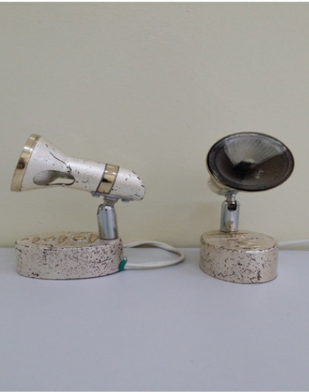
The purpose of this study is to assess the effect of long-range infrared radiation on endothelium-dependent vasodilation in patients with erectile dysfunction. Materials and methods. The study included 118 men aged 32-71 years with ED, as well as 20 men (control group) aged 28-66 years without ED. All men with ED underwent a comprehensive examination, which included the collection of a general medical and sexological history, a survey on the International Index of Erectile Function, a general examination, a study of hormonal status, blood lipids and glucose, as well as FGD using vasoactive drugs (papaverine 20-40 mg; alprostadil 5-10mkg). In addition, the study of the endothelial function of the cavernous arteries was performed in all patients. The function of the arterial endothelium was evaluated using ultrasonic examination (ultrasound) of the diameter before and after the use of long-range IR emitters (Figure 1). (ZB series registration certificate No. UZTT 00898 - working wavelength range of useful radiation 22.5 μm) according to our developed methodology. The experimental nature of the test was explained to all participants and consent was received from them to conduct it. Ultrasound of the cavernous arteries was performed using an “HD3” device from Philips (Holland).
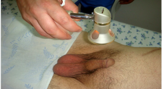
In the position of the patient on the back, a linear sensor L 9-5 was placed longitudinally on the ventral surface of the penis. The diameter of the cavernous arteries was measured as the distance between the opposite walls of the vessel and the location of the sensor was noted with a pencil. After determining the average diameter of the cavernous arteries of the penis, an IR emitter (ZB series) was exposed with an exposure of 3 minutes from the ventral side at a distance of 10-12 cm from the penis (Figure 2). After 3 minutes, an ultrasound scan was repeated to measure the diameter of the cavernous arteries. For the calculation, the largest diameter of the cavernous arteries was used. The percentage of increase in the diameter of the cavernous artery (PUDKA) was adopted by us as the main indicator for assessing the endothelial function of the cavernous arteries. This indicator was calculated by the formula:

Figure 1: Far-range infrared emitters.

lupinepublishers-openaccess-journal-urology-nephrology

Figure 2: Methods of exposure of IR emitter to cavernous arteries

lupinepublishers-openaccess-journal-urology-nephrology

PUDKA = 100% x (D2 - D1) / D1,
where D1 is the diameter of the cavernous artery before exposure to an infrared emitter;
D2 - diameter of the cavernous artery after exposure to an infrared emitter.

All studies were carried out by one specialist in ultrasound diagnostics. The study was carried out in the morning, before the study, patients were advised to refrain from smoking and taking medications that affect the cardiovascular system. The average values of this indicator were calculated for the control group and patients with arterial and non-arterial ED. Statistical analysis was performed using t-student criterion and Pearson correlation coefficient. For analysis, computer software Statistica 6.0 was used; p <0.05 was recognized as statistically significant.

Results

divided into 2 subgroups: subgroup I - non-arterial ED of 42 patients (peak systolic velocity of 30 cm / s or more); Subgroup II - arteriogenic ED of 76 patients (peak systolic velocity less than 30 cm \ s). A comparative analysis of the average PUDA values in patients with different forms of ED revealed that this indicator is statistically significantly (p <0.001) less in the group of patients with arteriogenic ED compared with the control group, as well as in comparison with patients with other forms ED. An analysis of the data made it possible for us to propose a threshold level of PUDA for distinguishing organic ED from other forms of erectile disturbances, which amounted to 30%. The sensitivity and specificity of this indicator relative to the results of FDG in the diagnosis of arteriogenic ED were 100% and 96%, respectively. In patients with arteriogenic ED there was a significant correlation (r = 0.58 p <0.05) with the results of FDG (peak systolic rate) and PUDK values. An analysis of the results showed that the proposed method reveals a paradoxical vasoconstriction of the cavernous arteries, which is not determined when using FDG. When conducting the proposed diagnostic method, in no case did we observe complications and adverse reactions.

Discussion

The present study shows that the use of long-range IR emitters in determining the endothelial function of the cavernous arteries using ultrasound is a highly informative method for the diagnosis of arteriogenic ED. The developed technique for studying the endothelial function of the cavernous arteries has advantages over FDG and post-occlusion test, as it is a non-invasive method and technically simple to perform. It is known that the functional state of penile arteries depends on the biological activity of NO. Endothelial cells and nerve endings of non-cholinergic nonadrenergic neurons are sources of NO in the cavernous tissue of the penis. The synthesis of NO is carried out as a result of the action of enzymes of endothelial and neuronal NO synthase [14,15]. In our opinion, endothelial and neuronal NO synthases are activated by the quantum energy of the IR emitter, which leads to the release of NO. It is known that the quantum energy of radiation is inversely proportional to the wavelength, and if you take this into account, the effect on a person with radiation in the range of 8-9.8 μm (human own radiation is in the range of 9-10 μm) does not have negative side effects, since quantum energy of such sources does not exceed the quantum energy of the skin’s own radiation. The radiation of ceramic infrared emitters has a specific wavelength in the narrow spectral range. All types of emitters are characterized in that the energy spectrum of their exposure corresponds to or below the energy spectrum of human radiation. Emitters in these ranges have no effect on a healthy person, since it is transparent to them [16,17]. The paradoxical vasoconstriction detected by the proposed method is due to the presence of oxidative stress. Longrange infrared radiation stimulates the production of superoxide radicals (NO; O) by blood phagocytes and tissue macrophages, which enhances oxidative stress. This is not observed during FDG. Endothelial dysfunction is an early stage of atherosclerosis and is systemic in nature. Functional damage to most arterial vessels often proceeds without clinical symptoms; therefore, the timely detection of early stages of vascular system damage is relevant. In our opinion, the developed method for determining the endothelial function of the cavernous arteries is able to detect endothelial dysfunction at the level of functional disorders, which is not always determined with FDG.

Conclusion

Thus, our proposed method for studying the endothelial function of the cavernous arteries after exposure to a far-infrared emitter is highly informative in the diagnosis of arteriogenic ED, especially in the stage of functional disorders. This method will take its place in a comprehensive examination of patients with ED. Conflict of interest. The author claims no conflict of interest.

Acknowledgment

To the Head of the Clinical and Diagnostic Department of the Central Clinical Hospital No. 1 of the Main Medical Directorate under the Office of the President of the Republic of Uzbekistan, Doctor of Medical Sciences G. A. Rozykhojaeva.

https://twitter.com/lupine_online


Tuesday, September 8, 2020

Lupine Publishers | Culture and Sensitivity; What Is Behind

Lupine publishers | Journal of Urology & Nephrology Studies


Short Communication

One or two years before I were considering the test of culture and sensitivity C/S in all specimens including urine is the optimal test or the last measure act in reaching the diagnosis for any. Recently this idea had gone away completely not due to the unknown extent of false negative and positive activity of the antibiotics due to vivo and vitro affairs. It is due to a fact that the revealed bacteria are not the point of our mystery especially in chronic repetitive urinary tract infection which may end in renal failure. The bacteria mentioned in the result forma or paper are what I term them as ‘’ Bad face or Quarrelsome face’’ this means if someone with this adjective is a person who is sent by someone to another someone to e.g. collect a post-due dept the owner failed to have it in usual fashion, or better when a gang boss is behind the jail bars send his outside guys to perform actions planed by him. This later example is the most correct description to the fact I want to reach which is the bacteria in the C/S result are the extra-cellular bacteria ECB where as the real problem confined in the depth of the cells where the intra-cellular bacteria ICB reside. The cells of urinary tract lining or underneath. 

Take this for example ( however I said recently ) I started to realize it may be got mature or hundred percent sure after accumulation of data that this idea is a fact. The example is; around ten years ago me and some surgeons and physicians went as a team of experts to some country to correct some health issue, three days post arrival one of our team which is a consultant urosurgeon asked for return home, I asked him what the cause could be, he replied with sadness that his mother suffer from chronic urinary tract infection and she is now tired so he need to find some solution especially it is resistant to recover. Really I laughed reflexly and told him we come to this country as expert to solve their problems while your mother had chronic UTI and you cannot put an end for it! He made no other response other than the first by saying in sad manner and his head inclined to the earth ‘ that is right I cannot help my mother further’ then me as a friend try to help asked him ‘’ did you do C/S” he said yes, what was the result ? he replied E. coli. I commented that he should consider Brucellosis to treat his mother with because Brucella is the ICB which sit inside the lining cells and make certain environment encourages the growth of certain biotic life and here we find E. coli as a “ bad face” to mis-lead and direct our attention away from it. Certain environment conditions make a given growths like a damp room find into it spider webs which keeps increase as time passes. 

This consultant urosurgeon did not make any objection on my instruction and I felt some comfort from his side as if I gave some exit however I am neurosurgeon and were not a consultant at that time. From where this principle came for me! More than twenty years ago I adopted the digging into the biological basis of neurosurgical pathologies. At the beginning it was based on clinical trial treatment after pointing the that Brucella what is behind the symptomatology and hence the pathology behind clinical condition ( lab is not reliable as serological test where if negative all what we do is missing the patient’s real problem to go to palliative and symptomatic measures) the results in my field were excellent starting with spine problems like low-back pain. As time and positive results pass on a new facts spring on earth like some other health problems that were accompany the treated issue faiding out to show in analysis the main issue and these faiding other health problems are mere pictures or more precisely are complications to the main health problem which in end one realizes it is a chronic or sub-acute bacterial systemic infection. As time passes more and more organs and tissues become involved. Lately PCR open tissue examination for Brucella proved that this idea is a fact where the results at the beginning were 25% positive for Brucella in my patients when the tissue where taken from Sacroiliac joint. Then around 60% positive when Trapezius muscle open biopsy was considered. By admitting screen molecular test with Micro-array and I enlisted around fifteen ICB now for direct sampling like spine and brain and so for my cases which biologically shows the other pathologies share similar. Urinary bladder cystoscope or renal biopsies for PCR screen may reveal this fact either. M. tb. And others are interpretation difficulty for me now where they emerge on my field.

https://twitter.com/lupine_online

Monday, September 7, 2020

 Lupine publishers | Hypertriglyceridemia and Remodeling of the Left Ventricle in Patients with Chronic Kidney Disease

Lupine publishers| Journal of Urology & Nephrology Studies

Abstract

Aim: The aim of the study was to study the clinical and functional features of renal dysfunction in conditions of hypertriglyceridemia and remodeling of the left ventricle.

Materials and Methods: 176 patients with chronic kidney disease (CKD) were examined, 111 of them with hypertriglyceridemia (HTG) in association with left ventricular hypertrophy (LVH) 1st group and 65 patients with LVH in the absence of HTG, matched by gender and age. Along with general clinical studies, all patients were assessed for lipid spectrum parameters, measurement of cystatin C in blood plasma, and indicators of arterial stiffness (augmentation index and stiffness) and echocardiography were analyzed.

Results: In the 2nd group (HTG + LVH), the number of patients with type 2 diabetes mellitus (DM), a combination of hypertension, coronary heart disease and gout were significantly higher (p<0.05). Persons with chronic obstructive pulmonary disease, chronic pyelonephritis and cerebrovascular diseases were significantly more common in the 1st group (p <0.05). In the 2nd group, the value of BMI [(30.4 ± 5.2 kg/m2 against 28.1±5.7 kg /m2; p = 0.013), the level of systolic blood pressure (BP) (140 ± 19 mm Hg). v. 134 ± 18 mmHg; p = 0.042), thickness of the posterior wall of the left ventricle (LV) (0.98 ± 0.18 cm versus 0.90 ± 0.16 cm; p = 0.008), relative thickness LV walls (0.385 ± 0.107 units versus 0.357 ± 0.060 units; p=0.032), the number of patients with concentric LV hypertrophy (29.2; versus 13.5%; p=0.008), total cholesterol content (5.80 (4.87;6.80) mmol/l vs. 4.75 (4.0;5.38) mmol / l; p=0.000); lowdensity lipoprotein cholesterol (3.56 (3.12;4.63 ) mmol/l versus 3.09 (2.61;3.79) mmol/l; p=0.045) cystatin C (1.25 (1.08;1.70) mg / l versus 1.16 (0.99;1.42) mg/l; p=0.026) turned out to be significantly higher, and the calculated glomerular filtration rate (eGFR) significantly lower (57.0 ± 22 ml / min versus 65.0±23 ml/min; p=0.028)] compared with the 1st group. A positive correlation was noted between the level of central BP (r=0.264; p=0.003), the augmentation index (r=0.224; p=0.011) and plasma cystatin C (r= 0.486; p=0.000) with the value of the indexed LV myocardium mass (LVMI) in the total sample surveyed. A negative correlation was recorded between the eGFR and LVMI (r=-0.425; p=0.000).

Conclusion: In the presence of hypertriglyceridemia in patients with left ventricular hypertrophy, cystatin C increases in blood plasma and GFR decreases with a tendency for augmentation index to increase.

Keywords: Hypertriglyceridemia; Left Ventricular Remodeling; Chronic Kidney Disease; Cystatin C

Introduction

The number of patients with chronic kidney disease (CKD) is increasing annually, which is becoming an important medical and social problem of our time [1,2]. Patients with CKD have an increased risk of developing cardiovascular diseases (CVD), so they should be examined to identify these complications [3]. Hypertriglyceridemia (HTG) can realize its effect on the progression of renal damage through the development of intrarenal atherosclerosis and through the toxic effect of lipid particles on nephron structures [4]. It was shown that already in the early stages of CKD, the plasma TG level rises, the highest TG levels are detected in patients with nephrotic syndrome and in patients receiving renal replacement therapy (RRT) [5]. This is associated with a decrease in the activity of lipoprotein lipase enzymes and triacylglycerol lipase in the liver. Under conditions of HTG in renal dysfunction, the production of proinflammatory cytokines increases and proteinuria is aggravated, which contributes to the progression of CKD and an additional increase in cardiovascular risk [6,3]. A decrease in GFR induces the development of left ventricular hypertrophy (LVH), the frequency and severity of which determine the prognosis for CKD [7]. Structural and functional changes of the heart in CKD primarily, hypertrophy and change in the geometry of the LV, on the one hand, are considered as a complication, and on the other, as a factor in the progression of renal dysfunction [8-10]. The combination of HTG and LVH in the general population of people has been studied sufficiently. However, there are few works where the value of HTG as an independent risk for renal dysfunction and CVD would be considered. This dictates the need to improve the early diagnosis of heart and kidney damage in HTG. In this regard, the study of LV restructuring and the functional state of the kidneys among persons with HTG is of considerable interest.

Materials and Methods

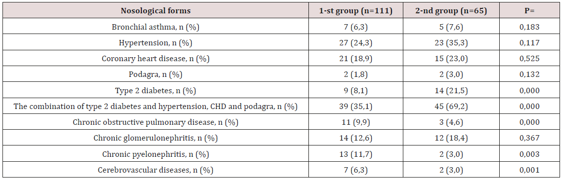
The work was performed in the clinical departments of faculty therapy of the KSMA n.a.I.K. Akhunbaeva and therapy №2 KRSU n. a. B.N. Yeltsin, Bishkek. The study included 176 patients over the age 18 with an established diagnosis of CKD. The midddle age of the examined individuals was 52.9±13.3 years. Diagnosis of hypertriglyceridemia [11], left ventricular hypertrophy [12] and CKD [13] were carried out in accordance with existing standards and recommendations. The study was approved by the Ethics Committee of the KSMA n.a. I.K. Akhunbaeva. Nosological characteristics of the patients included in the study are presented in Table 1. The study design is a momentary case-control study. The criteria for inclusion in the study was the presence of echocardiographic (EchoCG) signs of LVH and CKD. The study did not include individuals with endstage CKD who are on renal replacement therapy (RRT), or patients with valvular heart disease. All the examined individuals were divided into two groups depending on the TG content of the blood plasma. Group 1 (n = 111) included patients with a plasma TG level < 1.7 mmol/l, the 2nd group (n=65) with plasma TG ≥ 1.7 mmol/l, i.e. with the presence of GTG. Physical examination included an assessment of the general condition, a clinical measurement of blood pressure (BP) in both hands in a sitting position using the Korotkov method, a was determined body mass index (BMI), was calculated and the number of heartbeats (HR). BMI was calculated using the formula: BMI = body weight (kg)/height2 (m2).

Table 1: Clinical and anamnestic characteristics of research patient.
lupinepublishers-openaccess-journal-urology-nephrology

Note: CHD - coronary heart disease; Type 2 diabetes; n- is the number of patients; p-credibility.

The laboratory study included an assessment parameterof lipid spectrum s (total cholesterol (cholesterol), low-density lipoprotein cholesterol (LDL cholesterol), high-density lipoprotein cholesterol (HDL cholesterol) and TG), uric acid, calcium and fibrinogen in blood. Additionally, all patients were determined by the concentration of cystatin C in plasma by the immunoturbidimetric method. The severity of renal dysfunction was determined on the basis of the GFR, calculated according to the F.J. Hoek formula: GFR = 80.35 / Cys-4.32 [14]. The instrumental part of the work consisted of studying the parameters of arterial stiffness and augmentation (Alp, augmentation index) using the Angioscan-01 device (AngioScan Electronics LLC, Russia) [15]. Transthoracic EchoCG with simultaneous tissue myocardial dopplerography was performed on a Vivid Q ultrasound scanner (USA, 2014) according to the standard technique. The thickness of the walls, the dimensions of the left ventricular cavity (LV), the diameter of the exit aorta (cm) and left atrium (cm) were estimated from parasternal access along the long axis of the LV. Measured the thickness of the interventricular septum (IVS, cm) and the posterior wall of the left ventricle (LV, cm) in diastole, the final diastolic (FDM, cm) and the final systolic dimensions (FDM, cm) of LV were determined.

LV systolic function was estimated by its ejection fraction (EF, %), which was calculated using the formula L.E. Teichholtz (1976) in the absence of zones of hypo- and akinesis [16]. In order to assess the diastolic function of the left ventricle in the mode of pulsed Doppler sonography, the transmitral diastolic flow rate (E / A) and the time of blood flow slowing down of the early diastolic filling of the left ventricle (DT) were measured. The LV myocardium mass (MLMH) was calculated by the formula R.B. Devereux et al [17]. (1986): MLM (g) = 0.8 - {1.04 - (KDR + MZhP + ZSLZH) 3 - KDR3} +0.6 [17]. The LV myocardial mass index (LVMH) was defined as the ratio of LVML to body surface area. The criteria for LVH and types of LV myocardial remodeling were determined in accordance with the recommendations of ESC from 2013 [12]. For the assessment of LVHL, the LVMI was calculated, the upper value of which was 95 g/m2 for women and 115 g/m2 for men. The relative wall thickness (RWT) of the LV was calculated for each patient as (IVM + LVLS)/ LV CRD. For an increase in RWT, was taken a value of more than 0.42 [12]. The criteria of concentric and eccentric variants of LVHL were considered to be LVMI values greater than normal, RWT> 0.42 and LVMI higher than normal, but RWT <0.42, respectively. Statistical analysis of the data was carried out using the software package “Statistica 10.0”. All quantitative variables are presented as mean ± standard deviation, medians and quartiles [Me (Q25; Q75)]. Significance of differences between groups was assessed using t-Student test (for variables with a normal distribution) and the Mann-Whitney test (for variables with a non-parametric distribution) [18]. Correlation analysis was carried out by the Pearson criterion - for variables with a normal distribution and the Spearman coefficient (for variables with a non-parametric distribution). For all types of analysis, p <0.05 were considered statistically significant.

Results of Research

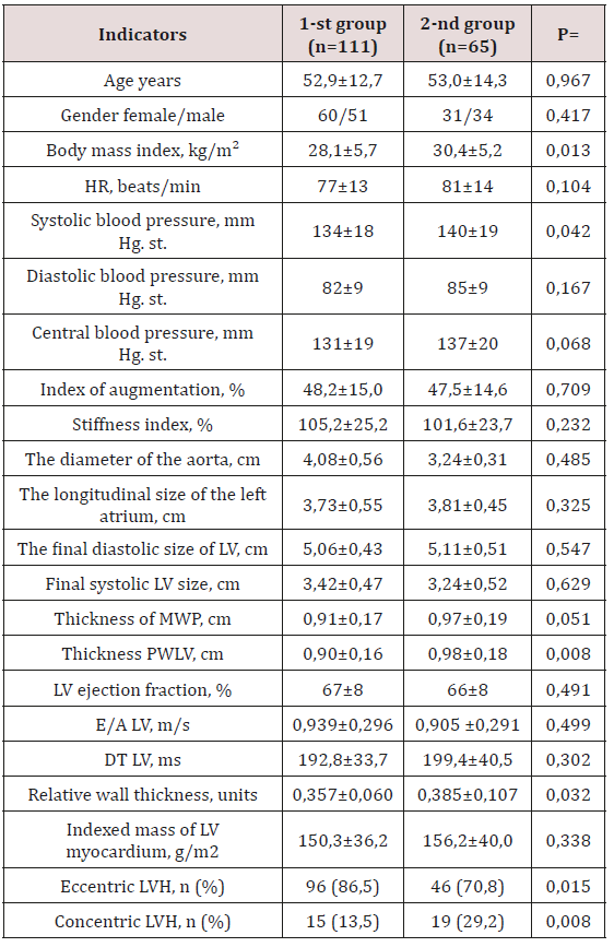
The distribution of patients with LVH without HTG and with HTG is presented in Table 1. Among the examined groups with HTG, the proportion of patients suffering from type 2 diabetes mellitus (DM) and its combination with hypertension, coronary disease, and gout was significantly higher in the 2nd group compared with the 1st group. In contrast, individuals with chronic obstructive pulmonary disease (COPD), chronic pyelonephritis and cerebrovascular diseases (CVD) were significantly more common in the 1st group. When assessing the general characteristics of the studied groups, it was noted that the initial patients were similar in age and sex (Table 2). In the 2nd group, i.e. in patients with HTG, the value of BMI was significantly higher (30.4±5.2 kg/m2 against 28.1±5.7 kg/m2; p=0.013) compared with the 1st group. Analysis of hemodynamic parameters showed a higher level of systolic blood pressure (140±19 mm Hg versus 134±18 mm Hg; p=0.042) in the group of individuals with HTG compared with the 1st group. It should be noted that there were no intergroup differences in heart rate, diastolic and central blood pressure, augmentation index and stiffness (Table 2). According to EchoCG, the diameter of the output aorta, the end systolic and diastolic size of the LV, the longitudinal size of the left atrium, the LV ejection fraction and LVMLI did not differ significantly. There was a tendency to increase the thickness of the IUP in the group of persons with HTG. At the same time, in this group there was a statistically significant increase in the SLWL thickness (Table 2). All examined individuals in both groups had similar LVML, E/A and DT LV values. Significant differences between patients of the two compared groups were determined in terms of LV RWT. As can be seen from Table 2, the RWT of LV patients of the 2nd group significantly exceeded the RWT of the LV patients of the 1st group (p=0.032).

Table 2: Clinical - laboratory parameters of the examined patients with CKD.
lupinepublishers-openaccess-journal-urology-nephrology

Note: n- is the number of patients; p - reliability; HR - heart rate; BP - blood pressure; LV - left ventricle; LVH - left ventricular hypertrophy; DT is the flow deceleration time.

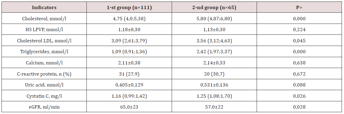
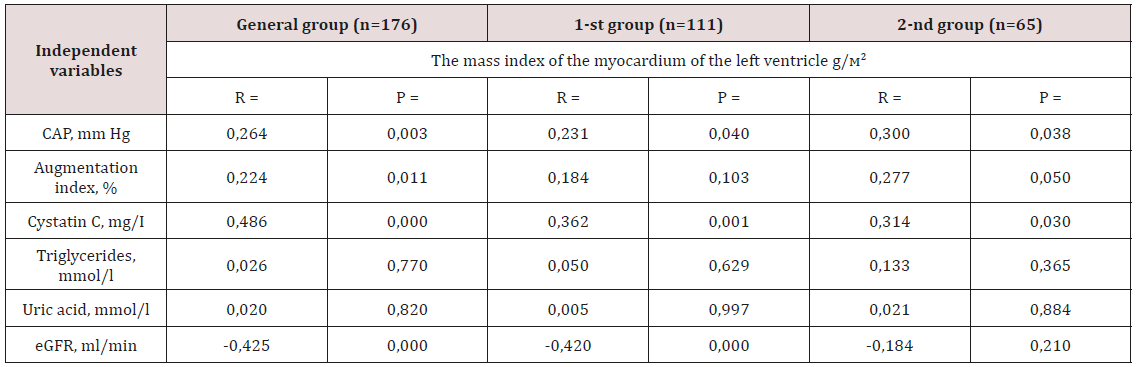
Analysis of the frequency of occurrence of types of LV restructuring of the two compared groups showed a significant difference in values (p<0.05). Thus, the proportion of patients with a concentric type of LVH was significantly higher among patients with HTG (29.2% vs. 13.5%; p=0.008), and the eccentric type significantly prevailed in the 1st group (86.5% vs. 70.8 %; p=0.015). At the next stage of the study, were analyzed the biochemical parameters of the examined groups, the results of the analysis are presented in Table 3. As was to be expected, in the group of individuals with HTG, the median total cholesterol, TG and LDL cholesterol was significantly higher (p <0.05). The calcium content and the number of patients with an increase in CRP in the blood plasma were equivalent in both groups. A clinically significant increase in plasma uric acid levels was recorded in the 2nd group. At the same time, in the same group of patients, there was a significant increase in plasma cystatin C content (p<0.05) and a decrease in calculated GFR (p<0.05). Evaluation of the relationship between the value of LVMI and clinical and laboratory parameters was carried out first in the general and in each group separately (Table 4). The presence of a reliable positive correlation relationship was obtained between the level of central BP (r=0.264; p=0.003), the augmentation index (r=0.224; p=0.011) and the concentration of cystatin C in the blood plasma (r=0.486; p=0.000) total sample. Feedback was observed between eGFR and LVMI (r=- 0.425; p=0.000). Similar correlation shifts with the exception of the augmentation index also occurred in the 1st group (Table 4). In the 2nd group, a direct close direct connection was found between the content of cystatin C in blood plasma, the level of central BP with the value of LVMI.

Table 3: Parameters of biochemical analysis of blood of the examined groups.
lupinepublishers-openaccess-journal-urology-nephrology

Note: eGFR - estimated glomerular filtration rate. HDL cholesterol - high density lipoprotein cholesterol; LDL cholesterol - low density lipoprotein cholesterol; number of patients.

Table 4: Correlation analysis between clinical laboratory parameters and the value of LVMI in two groups.
lupinepublishers-openaccess-journal-urology-nephrology

Note: CAP - central arterial pressure; eGFR - estimated glomerular filtration rate; p - reliability; n is the number of patients; r is the correlation coefficient.

Discussion

Despite advances in the study of diseases underlying CKD, the prognosis for this category of patients remains unfavorable. The concentration of TG-rich lipoproteins increases in the blood plasma at the early stages of CKD even at normal values of creatinine [19]. It should be noted that HTG is the most frequent variant of dyslipidemia in patients with kidney pathology [20,21]. A combination of HTG and an increase in the LV myocardium mass with a change in its geometry is believed to be associated with a further decrease in renal function and the risk of mortality, especially in the terminal stages of CKD [22,23]. It is important to note that HTG in patients of therapeutic profile has a double meaning. First, HTG is an important factor in the development and course of multifocal atherosclerosis and related cardiovascular complications. Secondly, an increase in the level of TG is directly related to the risk of developing renal dysfunction [20,21,24]. However, high TG levels are often combined with low HDL cholesterol levels and high LDL cholesterol levels. This fact is completely consistent with our results, i.e. in the group of patients with HTG, there was a statistically significant increase in the concentration of total cholesterol and LDL cholesterol (Table 3). Some meta-analyzes of TG are identified as an independent risk factor for CVD [24,25]. Moreover, recent genetic studies have confirmed the view that elevated TG is a direct cause of CVD development [26,27]. According to our data, among patients with HTG, the average eGFR was significantly lower (Table 3). It was established that a slight decrease in GFR is reflected by changes in the concentration of cystatin C in the blood [28].

Serum cystatin C levels are considered as a sensitive marker of the severity of renal dysfunction and the risk of LVH [29]. In studies A. Shankar et al. (2011), it has been shown that higher levels of cystatin C in individuals with CKD are associated with arterial hypertension [30]. In our study, we were also able to demonstrate a positive correlation between the content of cystatin C in blood and the value of LVMI in all groups (Table 4). The results of our work are consistent with data from other studies that have shown a relationship between elevated levels of cystatin C and LVH [31,29]. In persons with CKD, LVH is much more common, and the life expectancy of a patient with renal dysfunction and left ventricular hypertrophy is significantly reduced [32]. HTG also contributes to the occurrence of LVH in patients with CKD [20]. The presence of HTG stimulates the formation of adhesion molecules, the adherence of leukocytes to the surface of the endothelium, increases the secretory activity and proliferation of macrophages in the atherosclerotic plaque, activates the migration and proliferation of smooth muscle cells. A consequence of these effects is the development of endothelial dysfunction, increased stiffness of the vascular wall and myocardium [20,21]. Atherosclerotic changes in the arterial bed lead to ischemia of cardiomyocytes, activation of necrosis and apoptosis [33,34]. The decrease in the number of functioning cardiomyocytes, interstitial remodeling is accompanied by the development of both systolic and diastolic myocardial dysfunction [35].

We discovered a direct relationship between the magnitude of the augmentation index and the presence of LVH in the general group. A direct relationship was established between the increase in the augmentation index and the level of systolic blood pressure [36]. Was shown a close relationship of the blood lipid spectrum with vascular stiffness indicators [37]. The increase in the stiffness of large arteries plays an important role in the pathogenesis of many CVDs and, above all, in the development of hypertension [38,39]. On the other aspect, an increase in blood pressure causes changes in the vascular wall, incl. with the development of arteriosclerosis, which can lead to an increase in arterial stiffness of the renal arteries [40]. The augmentation index is a surrogate indicator of arterial stiffness and determines the state of the vascular bed from the central arteries to the microvasculature [41]. As a result of an increase in the augmentation index, the load on the LV increases, which certainly leads to the development of LVH [42,43]. Concentric hypertrophy is the most unfavorable type of remodeling, which is associated with the greatest number of complications [44-46]. The role of LVH and its relationship with the clinical and laboratory manifestations of CKD at the predialysis stage continue to be studied. According to the results of our study, HTG and LVH were reliably accompanied by an increase in systolic blood pressure, BMI, cystatin C, LDL LDL-C and a decrease in GFR. The negative impact of these clinical and laboratory parameters on the LV myocardium was manifested by an increase in the number of patients with a concentric type of LVH.

Conclusion

Hypertriglyceridemia in patients with left ventricular hypertrophy is accompanied by an increase in blood cystatin C, a decrease in GFR, and a tendency for augmentation index to increase. 

For More: Lupine Publishers Open Access Journals Please visit our Website:
https://lupinepublishers.us/

https://twitter.com/lupine_online


Radiology; USG and Colour Doppler of Post Renal Transplant Complications

  Abstract Kidney transplant is the treatment of choice for patients with end-stage renal disease. Kidney transplant offers better...