Lupine Publishers | Journal of Clinical Gastroenterology and Hepatology: is a multi-faceted Open access online journal exclusively allocated to novel work in the fields of Gastroenterology and Hepatology: By Lupine Publishers
https://lupinepublishers.us/
Showing posts with label journal of Urology & Nephrology. Show all posts
Showing posts with label journal of Urology & Nephrology. Show all posts
Introduction: Female urethral stricture is a highly
under-reported and underdiagnosed condition encountered by the
reconstructive urologist. Urethral dilatation is often performed with
urethroplasty offered in select cases. In the present study, we
describe our results in a series of women surgically treated for female
urethral stricture disease using a suprameatal approach with
a buccal mucosal graft dorsal on lay technique. Materials and Methods: All females diagnosed of urethral stricture who underwent buccal mucosal graft urethroplasty from
January 2015 to January 2020 were evaluated retrospectively. Intraoperative and postoperative parameters were assessed. Results: A total of 14 female patients underwent buccal mucosal
urethroplasty were evaluated. The mean age of the patients
was 49.5 years ranging from 35 to 64 years. Mean preoperative maximum
flow rate [Qmax] on uroflometry was 6.5 ml/second and
the mean residual urine 156 ml. All patients underwent uneventful buccal
mucosal graft dorsal on lay technique. At 3 months follow
up, the mean Qmax was 23.2 ml/second with mean residual urine of 14 ml. A
Self-reporting satisfaction scores using the Patient
Global Impression of Improvement showed that seven patients scored 1
(very much better), four scored 2 (much better), two
patients scored 3 (a little better), and one scored 4 (no change) none
of the patients scored a 5 (worse).No recurrence was noted. Conclusion: Buccal mucosal graft urethroplasty is a feasible surgery for female urethral strictures with minimal short term
complications.
Introduction: Peritoneal dialysis (PD) and haemodialysis (HD)
are two complementary and non-competitive renal replacement
therapy (RRT). A patient can be transferred from one technique to the
other. The objective of this study was to assess the reasons for
transferring patients from PD to HD and to follow their outcome. Patients and Methods: This is a 16-year descriptive and
analytical retrospective study (March 1, 2004 - August 31, 2020)
conducted at the PD unit of the Aristide Le Dantec University Hospital
in Dakar. Were included, patients on PD for at least 30 days,
over 18 years of age and permanently transferred to HD. The probability
of survival for any duration of post-transfer follow-up was
estimated by the Kaplan-Meier method. Results: The analysis covered 98 out of 113 cases. The mean age
of the patients was 45.2 ± 14.09 years at the initiation of PD
and 47 ± 13.91 years at the time of transfer, with a sex ratio of 0.66.
The mean duration in PD was 19.9 ± 17.25 months [range,
1.0-90.0 months]. The transfer to HD concerned 73.5% of patients in the
first two years. The reasons for transfer were mainly
associated with infection (82.7%), mechanical complications (23.5%),
social reasons (12.2%) and inadequate dialysis (6.1%). It
was programmed in 11.4% of cases and 6% of patients had a permanent
approach. At the endpoint date, the mean duration in
hemodialysis was 43.3 months with 42.8% of patients still in HD. There
was a kidney transplant patient; a return to PD. Mortality
was 34.6%. The mean HD survival was 126 months. There was a
statistically significant relationship between infection as a reason
for transfer and mortality (p = 0.047). Conclusion: The main reasons for transferring PD to hemodialysis identified in the literature are found in our context. This
transfer must be anticipated to reduce morbidity and mortality.
Background: Penile schwannoma is a rare tumor. They commonly present as an asymptomatic, painless and slow growing
mass. Other presentations include sexual dysfunction, most commonly dyspareunia, followed by erectile dysfunction, abnormal
penile curvature or pain with ejaculation. Case presentation: A 26-year-old male presented atypically with painful nocturnal penile tumescence, along with multiple
nodules over the dorsal penis. Excision of multiple penile tumors under general anaesthesia was performed and histopathologic
examination revealed benign schwannoma. Conclusion: Our hypothesis is that the schwannoma lies along the
axis of the dorsal penile nerve, and compression of this nerve
occurs during his erection causing pain. However, there are limited
presentations of painful erections in penile schwannomas, and
we hope that future studies can help confirm this theory.
This pictorial essay is an educational article aiming to provide both
textual and visual portrayals consisting of a collection of
images and texts on an important issue by reviewing and extrinsic
encasing pathology of the ureter to provide a guide to those who
are involved in diagnostic intervention. While considering diseases of
the urinary system, physicians mainly focus on the kidneys
and the bladder. Only scant attention is paid to the ureters. Most of
the UTO due to calculi are readily identifiable whereas many
cases of ureteric exterior encasement are frequently missed from early
detection even by experienced clinicians and radiologists.
Failure in recognition of the encasement of ureters and its causes may
lead to mistaken diagnosis with resultant inappropriate
management. However, problems with the ureters can adversely affect the
functioning of the kidneys and could even be lethal. In
this article we focus only ‘encasement of ureters’ with a few common
examples and salient signs that help in the diagnosis.
In 1970 I started to investigate the Laser-Technology for urologic
surgery together with H. Müßiggang. First of all, it was to check the
interacting of the different lasers with biological tissue. The result
you can gather from the Figure 1 Decisive are the two factors:
absorption and scattering of light into the tissue. The strong light
absorption of the CO2 – laser leads to an excellent incision effect with
low edema reaction. The light of the is mainly absorbed in the tissue
by hemoglobin and pigment colorings and therefore suitable for the
destruction of highly vascularized tumors or malformations. For
achieving greater volume effects, - necessary
for destruction of solid tumors, bilharzial bladder-lesions and inflamed
areas in interstitial cystitis, - the Nd:YAG – laser was used by us
since 1976 (Figure 2). Presupposed for the clinical application of
lasers was the developing of a quartz glass fiber transmission system by
Nath, a physicist from the Neuherberg Laser Labor (1973) and the
developing of a special cystoscope insert, designed by my working-group
(Staehler, Frank et al.) and constructed by the Storz
Compagny/Tuttlingen/Germany (Figure 3). The next steps were the laser
induced shock wave lithotripsy, developed between 1978 to 1986
(Munich/Lübeck) and the photodynamic procedures for early tumor
diagnosis (Figures 5a & 5b).
Chronic kidney disease (CKD) is a leaping up public health matter. It
has an augmented effect on cardiovascular diseases and
affects every system in body. In developed countries the extent of
prevalence is available due to renal registries. In middle or
low socio economic countries the proportion of this evolving health
issue is not known. CKD is rising and a major contributor is
soaring number of diabetes mellitus worldwide. Other significant
addition in this cohort of CKD is by rising numbers of obese and
hypertensive patients. In order to curtail this rising health matter
immediate and intense measures both at preventive and curative
levels are required.
Aim: Due to lack of formal renal registry we wanted to see spectrum of chronic kidney disease (CKD) in our catchment area and
at what CKD stage they present to tertiary care hospital.
Methods: All patients who presented to Emergency department of
Multan Institute of kidney disease from 01 Sep 2017 till
Sep 2019 data were evaluated. Record of Emergency department patients
were taken from electronic system of our hospital. Some
patients had multiple visits and we took first visits kidney function in
our analysis. eGFR was calculated from serum creatinine with
help of CKD-EPI equation.
Results: Total 4303 patients were included in study. Males
were 60% and females 40%. Age range from 13 years to 96 year old.
945 patients were excluded as they were not falling in chronic kidney
disease category. Remaining 3358 patients had chronic kidney
disease. Sub-analysis according to CKD stage showed 66.17% patients
presented at CKD stage V.
Conclusion: Kidney disease is rising globally. Countries where
renal registries are established provide incidence of chronic
kidney disease ranging from 10 to 15%. Still a lot of countries
worldwide do not have established system of data collection so true
incidence is not established. Our work is first of its kind reported
from this area. Drastic preventive strategies are need of time from
health budget planners.
A lymphocele is a common finding after renal transplantation. The
majority of patients are asymptomatic. However, once a
lymphocele has become symptomatic, this condition has to be treated.
Lymphoceles may originate either from the lymphatic system
of the recipient or the transplanted kidney. The most sensible measures
to prevent their occurrence therefore seems to be to restrict
the transplant bed to the smallest permissible level with careful
ligature of the lymphatic vessels in the area of the kidney hilum.
Therapy of a lymphocele after renal transplantation should commence with minimally invasive measures and continue with
invasive procedures only if these are unsuccessful, namely, puncture and drainage then sclerotization, and then laparoscopic or
open marsupialization.
Objective: To describe step-by-step technique in Robot Assisted Radical Prostatectomy of our transperitoneal posterioranterior
technique for prostatic dissection with preservation of the endopelvic fascia, preservation of the puboprostatic ligaments
and dorsal venous complex.
Materials and Methods: Description of our surgical technique over 80 patients who underwent RARP and the characteristics
group from 2016 to 2019 excluding the rest of cases who went to different surgical approach and technique for robot-assisted
radical prostatectomy.
Results: The mean age was 63 years old, 7% of patients were
overweight and 7.5% had obesity. The mean pre-operative
prostate volume was 42.620 cc, mean prostatic specific antigen (PSA) of
10.414.8 ng/dl. The mean console time was 198±47.
The surgical margins were positive in 13.75% of the patients.
Complications were recorded in the peri-operative period, five (6.2%)
Clavien-Dindo I and six (7.5%) Clavien-Dindo II.
Conclusions: After 8 years of experience in our center we have
modified our technique of robot assisted radical prostatectomy,
improving our results, following different worldwide concepts in the
prostatic dissection. Even if necessary, to increase the number
of cases we have find an easier way to reproduce with acceptable
results.
Lupine publishers | Journal of Urology & Nephrology Studies
Short Communication
One or two years before I were considering the test of culture and sensitivity C/S in all specimens including urine is the optimal test or the last measure act in reaching the diagnosis for any. Recently this idea had gone away completely not due to the unknown extent of false negative and positive activity of the antibiotics due to vivo and vitro affairs. It is due to a fact that the revealed bacteria are not the point of our mystery especially in chronic repetitive urinary tract infection which may end in renal failure. The bacteria mentioned in the result forma or paper are what I term them as ‘’ Bad face or Quarrelsome face’’ this means if someone with this adjective is a person who is sent by someone to another someone to e.g. collect a post-due dept the owner failed to have it in usual fashion, or better when a gang boss is behind the jail bars send his outside guys to perform actions planed by him. This later example is the most correct description to the fact I want to reach which is the bacteria in the C/S result are the extra-cellular bacteria ECB where as the real problem confined in the depth of the cells where the intra-cellular bacteria ICB reside. The cells of urinary tract lining or underneath.
Take this for example ( however I said recently ) I started to realize it may be got mature or hundred percent sure after accumulation of data that this idea is a fact. The example is; around ten years ago me and some surgeons and physicians went as a team of experts to some country to correct some health issue, three days post arrival one of our team which is a consultant urosurgeon asked for return home, I asked him what the cause could be, he replied with sadness that his mother suffer from chronic urinary tract infection and she is now tired so he need to find some solution especially it is resistant to recover. Really I laughed reflexly and told him we come to this country as expert to solve their problems while your mother had chronic UTI and you cannot put an end for it! He made no other response other than the first by saying in sad manner and his head inclined to the earth ‘ that is right I cannot help my mother further’ then me as a friend try to help asked him ‘’ did you do C/S” he said yes, what was the result ? he replied E. coli. I commented that he should consider Brucellosis to treat his mother with because Brucella is the ICB which sit inside the lining cells and make certain environment encourages the growth of certain biotic life and here we find E. coli as a “ bad face” to mis-lead and direct our attention away from it. Certain environment conditions make a given growths like a damp room find into it spider webs which keeps increase as time passes.
This consultant urosurgeon did not make any objection on my instruction and I felt some comfort from his side as if I gave some exit however I am neurosurgeon and were not a consultant at that time. From where this principle came for me! More than twenty years ago I adopted the digging into the biological basis of neurosurgical pathologies. At the beginning it was based on clinical trial treatment after pointing the that Brucella what is behind the symptomatology and hence the pathology behind clinical condition ( lab is not reliable as serological test where if negative all what we do is missing the patient’s real problem to go to palliative and symptomatic measures) the results in my field were excellent starting with spine problems like low-back pain. As time and positive results pass on a new facts spring on earth like some other health problems that were accompany the treated issue faiding out to show in analysis the main issue and these faiding other health problems are mere pictures or more precisely are complications to the main health problem which in end one realizes it is a chronic or sub-acute bacterial systemic infection. As time passes more and more organs and tissues become involved. Lately PCR open tissue examination for Brucella proved that this idea is a fact where the results at the beginning were 25% positive for Brucella in my patients when the tissue where taken from Sacroiliac joint. Then around 60% positive when Trapezius muscle open biopsy was considered. By admitting screen molecular test with Micro-array and I enlisted around fifteen ICB now for direct sampling like spine and brain and so for my cases which biologically shows the other pathologies share similar. Urinary bladder cystoscope or renal biopsies for PCR screen may reveal this fact either. M. tb. And others are interpretation difficulty for me now where they emerge on my field.
Purpose: To study the possible association of cardiac function with morphological changes of the kidneys in glomerulopathies.
Materials and Methods: 55 patients with GP were examined (32 men, 23 women) aged from 17 to 58 (on the average 32.76 ± 10.3) years. Given the high prevalence left ventricular systolic diastolic dysfunction in patients with GP with impaired renal function, analyzed the data of patients with CKD stages 1-3. The mean GFR was 87.92 ± 28.2 ml / min / 1.73 m2. Histology, immunofluorescence and electron microscopic method of investigation were used in the study of nephrobiopsy data.
Results: Analysis of the frequency of morphological types of nephropathy in the examined patients made it possible to detect the prevalence of membranous glomerulonephritis (MG), which accounted for 43.6% of observations. IgA nephropathy was detected in 36.4% of cases. Focal-segmental glomerulosclerosis and minimal-change disease were much less common and in an equal proportion (7.3% of cases). Less often (in 5,4%) was marked by membrane-proliferative glomerulonephritis. Damage to glomerulus and endothelial proliferation significantly influenced the delay in the flow of early diastolic filling of the left ventricle deceleration and isovolumic relaxation time.
The Conclusion: The causes of the development of diastolic dysfunction of the heart are the damage of glomerulus, endothelial proliferation, deposits of the complement fraction C3 in GP.
Chronic kidney disease (hereinafter CKD) is an actual medical and social problem all over the world due to its significant increase in prevalence and adverse outcomes [1]. The mortality from cardiovascular diseases (hereinafter CVD) in patients with CKD is 15- 30 times higher than in the general population [2,3]. The achievements of modern nephrology have identified new approaches to the clinical analysis of complications of glomerulopathies (HP). A comparison of the CKD clinic with the frequency of complications showed that 74% of patients starting dialysis treatment are diagnosed with cardiac pathology [4]. So, in the work of Paoletti E [5] there is a decrease in survival due to an increase in the number of fatal and non-fatal complications in the presence of CVD in patients with chronic renal insufficiency. As for the pre-dialysis population, Weiner DE [6] analysis of four large studies (2,423 patients), with the definition of glomerular filtration rate (GFR) (15 to 60 mL / min / 1.73 m2) showed the dependence of the mortality rate on CVD in patients with CKD. In a study by Cai Q Z [7] significant changes in the side of deterioration of the cardiac structure were detected already in the first year of the disease in patients with CKD 3-5 stages. The authors explain this fact by the prolonged increase in the mass and volume of the left ventricle (LV) and the presence of diastolic (LVDM), rather than systolic LV dysfunction (LVDS). In the experimental work of Li K-L [8] it was found that maintaining normal diastolic function of the left ventricle in patients with CKD prevented the appearance of heart failure with reduced renal function. Modern methods of nephrobiopsy and improvement of immunomorfologic methods of analysis have opened the possibility to the study of live morphology of the kidney tissues more deeply, as well as to determine the role of the immune mechanism in the development of complications, including CVD [9]. Thanks to these data, the treatment of the disease, approaches to therapy and prevention of complications will change in the future [9]. However, at present, there are many unsolved issues in this area. There is still no clear correlation between clinical data and morphological manifestations of GP.
Objective: To study the features of the association of cardiac dysfunction with morphological changes in the kidneys with HP.
55 patients with GP were examined (32 men, 23 women) aged from 17 to 58 (on the average 32.76 ± 10.3) years. The glomerular filtration rate (GFR) was calculated by CKD-EPI (2013), and the CKD stages were determined according to NKF K / DOQI, Guidelines, 2002 [10]. Given the high prevalence of LVDS and LVDM in patients with CKD with reduced renal function, we analyzed the data of patients with CKD 1-3 stages. The mean GFR was 87.92 ± 28.2 ml / min / 1.73 m2. All patients underwent a complex of general clinical, biochemical, bacteriological, immunological, instrumental and morphological studies. General clinical methods included general analyzes of urine, blood and daily proteinuria. Echocardiography (ECHOKG) study was carried out according to the standard procedure on the “CX50 CompactXtreme Ultrasound System” with the identification of a complex of generally accepted morphofunctional parameters. To evaluate the diastolic function of the LV, a transmittal flow from the apical access in the 4-chamber section was examined, and tissue myocardial Doppler echocardiography.
In the study of nephrobiopsy, histological, immunofluorescent and electron microscopic methods were used. For the diagnosis of forms of GB, conventional morphological criteria were used [10]. The conclusion on the study of nephrobiopsy was established according to a single terminology in describing the pathological process and the morphological classification of the ICD X revision. In morphological analysis of glomerular changes the size of the glomerulus, the presence (degree) or absence of proliferation of mesangial cells and glomerular capsule epithelium, the state of the mesangial matrix and the structure of the basal membrane (thickening, uneven contours, splitting) are described. The algorithm of studying the kidney biopsy covered information on interstitial tissue (fibrosis, edema, infiltration) and the presence of adhesions (atrophy, dystrophy, necrosis). Morphological signs of the activity of the process included such processes as protein exudation in the lumen of the capsule, plethora of capillaries, plasmorragia, fixation on the basal membranes of glomerular capillaries and in the mesangium of immunoglobulins and fibrinogen. The statistical processing of the material was carried out using the Statistica 6.0 statistical software package. The results are processed using multivariate stepwise regression analysis. Differences were considered statistically significant at p <0.05.
In most patients (78.1%), the disease made its debut at the age of over 17, which is consistent with the literature data [11,12]. Clinically, most often there was a nephrotic syndrome (65.4%) and in most cases men (72.2%). Mean levels of systolic blood pressure (SBP) and diastolic blood pressure (DBP) were 130.3 ± 20.0 mm Hg and 83.2 ± 13.06 mm Hg, respectively. At the first stage of the study of our patients, the morphological characteristics of the GP is studied. Analysis of the frequency of various morphological forms of nephropathy in the subjects allowed to reveal the prevalence of membranous glomerulonephritis (MH), which accounted for 43.6% of observations. IgA nephropathy was detected in 36.4% of cases of primary HP. Among the subjects, focal-segmental glomerulosclerosis (FSG) and minimal-change disease (IMC) were encountered much more rarely and with an equal share (7.3% of cases). Less often (5,4%) was marked by membrane-proliferative glomerulonephritis (MPGN).
Light-optically glomerular changes were manifested in the form of minimal damage in 23.6%, segmental sclerosis in 41.8% and endothelial proliferation in 14.5% of cases. The occurrence of mesangial hypercellularity in the examined patients was 50%, the expansion of mesangium was 38.1%. In 52.7% of the patients, there were tubular injuries in the form of tubular atrophy, as well as interstitial fibrosis. Immunohistological examination of IgG was found in 43.6%, IgA - in 36.4% and IgM - in 20% of cases, and they had a granular distribution. Such a presence of deposits on the basement membrane of glomerular capillaries testified to the immune nature of kidney damage. Electron microscopy in 72.7% of cases recorded the merging of the legs of podocytes, and in 21.8% - mesangial expansion.
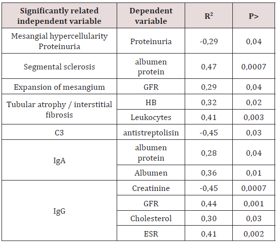
At the second stage of our work, we studied the factors that influence to the clinical picture of glomerulopathies. A multifactorial stepwise regression analysis was performed, where as independent variables included the indices of morphological changes in the nephrobioptate, as well as varieties of immunocomplexes. As dependent variables, the parameters of the general analysis and biochemistry of blood, as well as the echocardiographic parameters (systolic, diastolic function of the heart) were alternately included. We tried to analyze the relationship between the state of the filtration function of the kidneys and the immunological situation in the glomeruli. It turned out that in the general sample of patients, the presence of IgG (R2 = 0.44, P <.001) was a significant factor that adversely affected the value of RSKF. The presence of IgG in the composition of glomerular deposits, respectively, and reflected at the level of creatinine (R2 = -0.45, P <.0007). Reliable statistical results of our study indicate that the presence of immunoglobulins in the glomeruli reflects the severity of the nephritis. The ratio of histological changes and indices of proteinuria and blood tests are presented in Table 1.
Table 1: Morphological indicators, associated with clinicbiochemical sizes of blood.
Note: AP - albumen protein; GFR - glomerular filtration rate; Нв - haemoglobin; АСЛО - antistreptolisin; СОЭ – скорость оседания эритроцитов.
As follows from Table 1, there was a clear connection between the morphological pattern of changes in the renal parenchyma and a number of blood indicators reflecting the GP clinic. The effect of segmental sclerosis on the amount of serum protein was demonstrated by direct regression analysis (R2 = 0.47, P <.0007). To explain this situation, the following point of view seems appropriate: the detected changes in the glomeruli reflect significant damage to the glomerular filter, which create conditions for pronounced proteinuria and a change in the colloidal stability of the blood plasma [13]. Further, as can be seen from Table 1, the changes in mesangium included in the final model of multiple stepwise linear regression significantly influenced the calculated index of renal function (R2 = -0.29, P <.04). By analogy with the above, a reliable relationship was found when comparing mesangial hyper cellularity and proteinuria (R2 = -0.29, P <.04). Of no little importance was the study of the condition of the tubules. In general, we received evidence in favor of the presence of a relationship between tubular atrophy (TA), interstitial fibrosis (IF) and hemoglobin level in the blood (R2 = 0.32, P <.02). This phenomenon explains the fact that the severity of anemia in HH depends on the depth of damage of interstitial kidneys, since it is here that the formation of erythropoietins occurs.
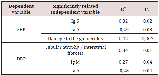
Table 2: Morphological variables associated with arterial pressure.
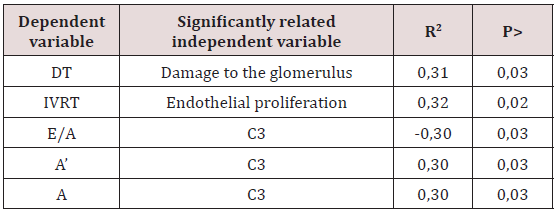
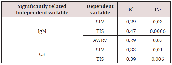
As for the ratio of immunohistology and blood pressure (Table 2), we found a significant relationship between the presence of TA / IF and DBP (R2 = 0.34, P <.01). It should be noted that in patients with TA/IF arterial hypertension (AH) was a consequence of tubulopathy, not nephrosclerosis. Our results are consistent with the data of Kotaro Haruhara et al. [14] who conducted a study of the relationship of tubulopathy and AH in patients with IgA nephropathy. We found the same pattern when comparing the parameters of damage to glomerulus and AH (R2 = 0.42, P <.002). The established dependence testified to the leading role of endothelial dysfunction in the development of arterial hypertension. This information is consistent with the data of Salmon AH et al. [15,16] who believe that proteinuria, as a consequence of damage of glomerulus, is considered a marker of endothelial dysfunction and glomerular hyperfiltration. At the same time, damage to glomerulus and endothelial proliferation (Table 3) caused a slowing of the time of blood flow of early diastolic filling of the left ventricle (DT) and isostolic LV relaxation (IVRT) (R2 = 0.31, P <.03 and R2 = 0.32, P < 0.02, respectively). Significant shifts in parameters of LV relaxation function were due to the appearance of complement fractions in the composition of immune deposits in capillaries C3. Therefore, the results obtained by us indicate an early renocardial interaction. Here, probably not final role-plays the presence of an inflammatory process, which leads to the development of diastolic dysfunction [17]. The pathomorphism of nephropathy is manifested by the development of systolic LV dysfunction [18] of great interest was the question of the effect of immune complexes on the architecture of the heart. Analysis of the parameters of our patients Table 4 showed a significant effect of IgM on systolic LV volume (SLV), thickness of interventricular septum (TMV) and the anterior wall of the right ventricle (PSP) (R2 = 0.29, P <.03, R2 = 0.47, P <.0006, R2 = 0.29, P <.03, respectively). Similar results were obtained in the study of the effect of complement on the heart. Thus, the dependence of the thickness of the walls of SLV, TIS on the C3 fraction of the complement fraction (R2 = 0.33, P <.01, R2 = 0.39, P <.006, respectively) has been elucidated. Consequently, the appearance of immune deposits under the capillary endothelium is systemic.
Table 3: Morphological features associated with parameters diastolic relaxation of the LV.
Note: DT - the time to slow the blood flow of early diastolic filling of the left ventricle; IVRT - the time of isovolytic relaxation of the left ventricle; E / A - the ratio of early to late diastolic filling; A’- late diastolic movement of the medial part of the mitral ring; A - late diastolic filling; C3 - complement.
Table 4: Immunomorphological findings associated with the parameters of the structure of the myocardium.
Note: SLV - systolic LV volume, TIS - thickness of the interatrial septum; AWRV - the anterior wall of the right ventricle.
a. Tubular atrophy and interstitial fibrosis are involved in the development of arterial hypertension in patients with HP.
b. Damage to glomerulus, endothelial proliferation, deposits of the complement C3 fraction in the vessels are presumably the cause of diastolic dysfunction.
c. The deposition of the IgM and C3 complement fraction in the capillaries are systemic and affect the size of the heart.
For more Lupine Publishers Open Access Journals Please visit our website
End Stage Renal Disease impacts the lives of over 725,000 Americans and their families. In 1972, despite the cost of supporting a rapidly growing chronic condition at the time, the United States initiated a system whereby everyone would be treated that qualifies for social security benefits, after a waiting period of three years (Medicare Secondary Payer was slowly extended over the years to 36 months after diagnosis as it currently stands), regardless of age, providing the platform for the debate of a potential nationalized health care system that currently seems to be in a period of uncertainty. However, over three decades through significant consolidation, the treatment of dialysis via two large provider chains (one fully integrated) have directed patients to hemodialysis in the out-patient setting without considering the technological or clinical benefits of home hemodialysis or peritoneal dialysis.
Doxorubicin is a hydrophilic anticancer drug. Due to hydrophilicity, it’s difficult to encapsulate in a hydrophobic core of nanomicelles. The main purpose of this study was to develop a hydrophobic ion paring complex (HIP) of doxorubicin using hydrophobic retinoic acid (Vitamin A). The resultant hydrophobic (DOX-RA) complex was utilized to prepare drug-loaded nanomicelles by co-precipitation method with penta block copolymer. Nanomicelles (DOX-RA/PBNM), was prepared by evaporation rehydration technique. DOX-RA complex was analyzed by H-NMR and FTIR. The nanomicelles were analyzed for size and zeta potential using dynamic light scattering (DLS) as well as transmission electron microscopy (TEM). The FT-IR and the H-NMR analyses confirmed the structures of the DOX-RA complex. Mean nanomicellar sizes were 25.5nm ± 5.00nm, and ζ-potential was approximately zero. H-NMR and FTIR analysis done on DOX-RA indicate peaks characteristic of both doxorubicin and retinoic Acid. This confirmed successful complex formation. Transmission electron microscopy (TEM) analysis revealed round shaped morphology and sizes similar to DLS results. In vitro release studies revealed that pentablock nanomicelles released Doxorubicin at a slow first order rate in phosphate buffer solution (PBS) at pH 7.4 compared to pH 5.5 and pH 4.0. Confocal microscopy analysis with DOX-RA/ PBNM indicated that pentablock nanomicelles were efficiently taken into prostate cancer (PC3) cells and doxorubicin was efficiently released from the nanomicelles into the cells. In addition, cell proliferation assay showed that nanomicelles ferried adequate amounts of Doxorubicin into PC-3 cells and inhibited cell growth significantly. Results confirm that DOX-RA complex facilitated the encapsulation of doxorubicin within nanomicelles increasing DOX intracellular concentration.
One or two years before I were considering the test of culture and sensitivity C/S in all specimens including urine is the optimal test or the last measure act in reaching the diagnosis for any. Recently this idea had gone away completely not due to the unknown extent of false negative and positive activity of the antibiotics due to vivo and vitro affairs. It is due to a fact that the revealed bacteria are not the point of our mystery especially in chronic repetitive urinary tract infection which may end in renal failure. The bacteria mentioned in the result forma or paper are what I term them as ‘’ Bad face or Quarrelsome face’’ this means if someone with this adjective is a person who is sent by someone to another someone to e.g. collect a post-due dept the owner failed to have it in usual fashion, or better when a gang boss is behind the jail bars send his outside guys to perform actions planed by him. This later example is the most correct description to the fact I want to reach which is the bacteria in the C/S result are the extra-cellular bacteria ECB where as the real problem confined in the depth of the cells where the intra-cellular bacteria ICB reside. The cells of urinary tract lining or underneath.