The Experience of Robotic Camera Holder During Laparoscopic Radical Retropubic Prostatectomy by Branimir Penev in Journal of Urology & Nephrology Studies (JUNS) - Lupine Publishers
Abstract
Background: Laparoscopic radical prostatectomy (LRP) is a challenging procedure for both the surgeon and the assistant(s). Frequently only one assistant is available who has to manipulate the camera and also assist with traction and/or suction. This may lead to poor image quality making the procedure more difficult for the surgeon. Previous robotic camera holders were bulky and expensive. We report on the impact of a new robotic camera holder on the outcome of extraperitoneal laparoscopic radical prostatectomy.
Methods: 72 patients undergoing LRP were prospectively analysed for length of surgery and blood loss. For the first 36 patients, the laparoscope was held by the first assistant while with the subsequent 36 cases, the Freehand® robotic camera holder was used. The assistant was at the same level of training over the study period.
Results: The introduction of the robotic camera holder significantly reduced the length of surgery by approximately one hour and blood loss by an average of 400ml.
Conclusion: The robotic camera holder significantly reduces operative time and blood loss.
Introduction
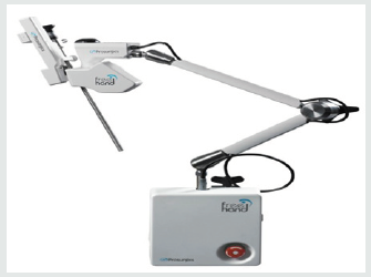
Laparoscopic Radical Prostatectomy (LRP) is a standard surgical approach for treating organ confined prostate cancer. The procedure has evolved significantly since it was first described by Schuessler et al. [1] in 1997 in their series of 9 cases. They used five 10-mm ports and took them an average of 9.4 hours. One of the most important aspects of laparoscopic surgery is the operator’s view of the surgical field. Provision of a consistently good view is paramount, which requires a skilled assistant to hold and manipulate the camera. Excessive camera movements, loss of orientation and communication problems with the assistant are troublesome and can potentially complicate the operation and jeopardise the outcome. In addition to camera holding, the assistant is often required to use their other hand for suction or retraction. The alternative is to have 2 assistants, but this is often not possible due to staffing issues. To aid the surgeon and assistant, camera holders have been developed to keep the camera steady. However, these devices were awkward and required repositioning for different views. Robotic camera holders have been used in the past but are expensive and cumbersome. Recently, a new robotic camera holder has become available. The FreeHand® (Prosurgics Ltd, Bracknell, Berks, UK) is a light weight robotic camera holder that is controlled by the surgeon’s head movement and a foot pedal (Figure 1). It is designed to facilitate surgery by improving image quality. This study describes our initial experience of the camera holder and assesses its impact on the learning curve of LRP.
Material and Methods
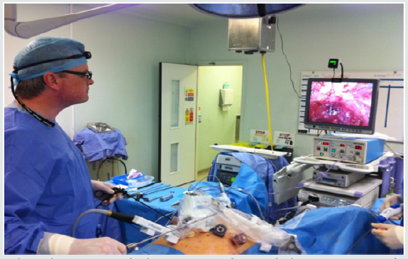
Seventy-two consecutive patients undergoing LRP were included in this study. The first 36 cases involved the camera being held by the assistant (human camera holding) and the subsequent 36 cases with the robot arm (FreeHand® camera holding). The operations were carried out between July 2010 and December 2012. The assistant was at the same level of training over the study period. Data was collected prospectively and analysed in 9-patient cohorts (8 cohorts in total) assessing operative time and blood loss. Operative time was recorded from initial skin incision to the application of dressings to the wounds at the end of the procedure. Blood loss was the total amount of fluid in the suction bottle minus the volume of irrigation fluid. All the cases were carried out by one surgeon (JD), representing his first independent experience in LRP. JD has contemporary experience in open prostatectomy and received appropriate training and mentorship in LRP. At the beginning of the procedure, and once the patient is under general anaesthesia on the operating table, the camera holder is attached to the side of the table. The arm of the device is covered with a sterile sleeve and then the patient is draped. Once all the ports have been placed, the robotic arm is attached to the laparoscope. The procedure was performed extraperitoneally using 4 ports - two 5mm and two 12-mm (one of which was the camera port). The endopelvic fascia, lateral and anterior bladder neck, ductus deferens, seminal vesicles and Denovilliers fascia were all incised to help mobilise the prostate. The dorsal venous plexus was then ligated and cut allowing the prostate to be excised. The bladder neck was then anastomosed to the urethra. The robotic arm provides a wide range of the movement with three axes of movement: left/right (180º horizontal arc), up/down (vertical tilt +70º to -20º relative to horizontal) and in/out (12cm zoom function). This allows the surgeon to have full control of the laparoscope. These movements are controlled by the head movements of the operating surgeon who wears a head-mounted infra-red controller device which transmits to an indicator unit at the top of the monitor (Figure 2). The movements only take place when the arm is activated by a footswitch to avoid unwanted movements. Comparisons were made between two consecutive cohorts using the Student’s t-test. Means were recorded with the standard error of the mean (95%CI). Statistical analysis was carried out using SPSS.
Figure 2: Theatre setup during laparoscopic radical prostatectomy showing the laparoscopic camera being held by the robot, the surgeon with head mounted controller and the indicator at the top of the monitor.
Results
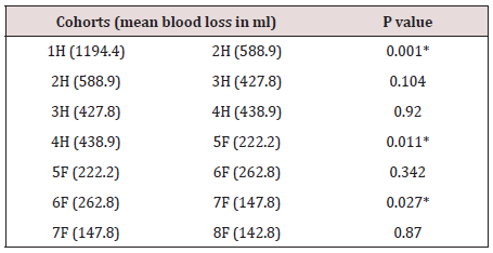
All the cases were performed laparoscopically and there were no conversions to open surgery. The device was safe, and setup took less than 5 minutes (after draping the patient as previously described). Patient demographics are shown in Table 1. There is no difference between the 2 groups with regards to age or prostate volume. Table 2 and Figure 3 show the mean operative times according to the cohorts. There was significant improvement in operating time for the first 3 cohort groups which levelled out for the 4th group. Following the introduction of the robotic camera holder, there was a significant mean reduction in operative time (Group 5) of 53 minutes. There was a further non-significant reduction in times (Groups 6 & 7) until a final significant drop in the last group. Table 3 and Figure 4 show the mean blood loss in the 8 groups. There was a significant reduction in blood loss at the beginning the series and following the introduction of the robot camera holder.
Figure 3: Box and whisker plot for the mean operative time ± standard error. H= Human camera holding; F=Freehand® camera holding.
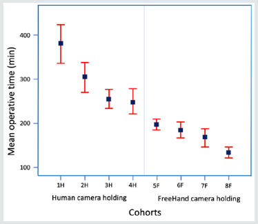
Figure 4: Box and whisker plot for the mean blood loss ± standard error. H= Human camera holding; F=Freehand camera holding.
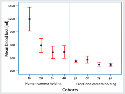
Table 2: Comparisons between the operative time means of two consecutive cohorts. H=human camera holding; F=FreeHand® camera holding. *significant.
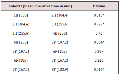
Table 3: Comparisons between the blood loss means of two consecutive cohorts. H=human camera holding; F=FreeHand® camera holding. *significant.
Discussion
The robotic camera holder is safe and offers stability and improvement in image quality during laparoscopic prostate surgery. It significantly reduces the length of surgery with a concurrent reduction in blood loss. This reduction in operative time benefits the patient and also offers a financial advantage. The learning curve of LRP varies amongst different surgeons, which is reflected in the published data; perhaps due to variations in their abilities, surgical techniques, previous laparoscopy experience and training. The learning curve for the operative time has been reported to be between 35- 150 cases [2-4] and blood loss is around 150 cases [2]. Our data suggest that the learning curve for blood loss and operative time appeared to improve in phases. Initially, the curve started to drop rapidly for the first 18 cases followed by a slow and insignificant decline. When the robotic arm was introduced at the 37th case, the curve then went down significantly and continued to improve slowly. In the last cohort, the mean operative time has significantly improved, which is perhaps explained by the change of our technique in the specimen retrieval. In the first 7 groups, the prostate was retrieved before the anastomosis between the bladder and urethra. This meant removal of one of the ports with loss of pneumoperitoneum, slight extension of the 12mm port access incision, removal of the specimen, replacement of the port secured with sutures and recreation of pneumoperitoneum. This was time consuming, and in the last group we changed our technique by retrieving the prostate at the end of the procedure resulting in a further saving of approximately 20 minutes. The use of robotic camera holding in laparoscopic urological surgery was introduced in the mid 1990’s. Partin et al. [5] concluded that it was safe and feasible to use a camera holder, which was controlled by a foot pedal, in various laparoscopic urological surgeries (excluding prostatectomy).
Various robotic camera holders have been introduced since, such as the AESOP (Automated Endoscopic System for Optimal Positioning), which is a voice-activated system.
The system requires individual setting for voice recognition and that each surgeon must have a voice card. Background noise interference could be an issue as well [6]. It is no longer available but cost approximately $84000 [7]. Another device is the EndoAssist, or the so called first generation robotic camera holder. It is a freestanding system that is controlled by a foot pedal and the operator’s head movements, in a similar way to the FreeHand® system. It has been successfully used in general surgery [8] producing good image quality. In a randomised trial, Aioni et al. [6] have managed to reduce the operating time for laparoscopic cholecystectomy from 74 to 66 minutes (p < 0.05) when using the EndoAssist compared to human camera holding. The disadvantage of the EndoAssist is that it is bulky, and being free- standing, it will not allow for a change of the patients’ position during the operation unless the device is disconnected and reset - which is time consuming. The FreeHand is more compact, cheaper and easier to set up than the EndoAssist. This second-generation system is attached to the side of the operating table, hence moves with the table, allowing the surgeon to reposition the patient at any time during the operation with ease. The system costs $20000 with $200 per case for disposables. The actual device is offered free of charge and the only cost is the disposables. This is offset by potential theatre time savings, on average of 1 hour per case. One of the other advantages of the robotic camera holder is that institutions that do not have the potential for experienced camera-holding assistants would probably benefit from use of the robotic camera holder. The question raised is does the Freehand® improve the outcome of LRP in terms of operative time and concurrent blood loss? The published data is still limited to one small randomised trial. Stolzenburg et al. [9,10] have compared 25 patients undergoing LRP with human camera holding versus 25 patients with robotic camera holding. They have concluded that the FreeHand provided a better image quality and with less camera error, but there were no significant differences in terms of operative time and blood loss. However, they also mentioned that the inclusion of a higher number of patients would have allowed them to provide more accurate comparative data for the two groups. In conclusion, robotic camera holding is safe and provides good quality, stable images. It reduces the operative time which in turn has financial advantages in cost savings.
References
- Schuessler WW, Schulam PG, Clayman RV, Kavoussi LR (1997) Laparoscopic radical prostatectomy: initial short-term experience. Urology 50: 854-857.
- Eden CG, Mischel G Neill MG, Louie-Johnsun MW (2008) The first 1000 cases of laparoscopic radical prostatectomy in the UK: evidence of multiple learning curves. BJUI 103: 1224 -1230.
- Rodriguez AR, Rachna K, Pow-Sang JM (2010) Laparoscopic extraperitoneal radical prostatectomy: impact of the learning curve on perioperative outcomes and margin status. Journal of the Society of Laparoendoscopic Surgeons 14: 6-13.
- Ghavamian R, Schenk G, Hoenig DM, Williot P, Melman A (2004) Overcoming the steep learning curve of laparoscopic radical prostatectomy: single-surgeon experience. Journal of Endourology 18: 567-571.
- Partin AW, Adams JB, Moore RG, Kavoussi LR (1995) Complete robotassissted laparoscopic urologic surgery: a preliminary report. J Am coll Surg 181: 552-557.
- Aiono A, Gilbert JM, Soin B, Finlay PA, Gordan A (2002) Controlled trial of the introduction of a robotic camera assistant (EndoAssist) for laparoscopic cholecystectomy. Surg Endosc 16: 1267-1270.
- Kraft B M, Jäger, Kraft K, Leibl B J, Bittner R (2004) The AESOP robot system in laparoscopic surgery: Increased risk or advantage for surgeon and patient? Surgical Ensosc 18: 1216-1223.
- Gilbert JM (2009) The EndoAssist robotic camera holder as an aid to the introduction of laparoscopic colorectal surgery. Ann R Coll Surg Engl 91: 389-393.
- Stolzenburg J, Franz T, Do M, Turner K, Liatsikos E (2009) Comparison of the FreeHand® robotic camera holder to human assistants during the endoscopic extraperitoneal radical prostatectomy (EERPE) - a prospective randomised study of 50 cases. J Endourology 23: A3.
- Jens-Uwe Stolzenburg, Toni Franz, Panagiotis Kallidonis, Do Minh, Anja Dietel (2010) Comparison of the Freehand® robotic camera holder with human assistants during endoscopic extraperitoneal radical prostatectomy. BJUI 107: 970-974.
To know more about Urology Nephrology Journal articles please click: https://lupinepublishers.com/urology-nephrology-journal/index.php
For more Lupine Publishers Open Access Journals Please visit our website: https://lupinepublishers.us/


No comments:
Post a Comment